Sensory input monitors stimuli inside and outside the body, Integration interprets sensory input, and Motor output activates effector organs to respond.
Central Nervous System (CNS)
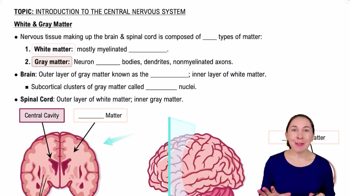
Consists of the brain and spinal cord; acts as the integration and command center of the nervous system.
Peripheral Nervous System (PNS)
Includes all neural structures outside the CNS such as cranial and spinal nerves, sensory receptors, and ganglia; connects CNS to the rest of the body.
Sensory (Afferent) Division of PNS
Carries impulses from sensory receptors to the CNS; includes somatic sensory fibers from skin and muscles and visceral sensory fibers from organs.
Motor (Efferent) Division of PNS
Transmits impulses from the CNS to effector organs like muscles and glands.
Structure of a Nerve
A nerve is a cordlike organ of the PNS made of peripheral axons enclosed by connective tissues: endoneurium surrounds axons, perineurium bundles fibers into fascicles, and epineurium surrounds the entire nerve.
Classification of Nerves
Nerves can be sensory (afferent), motor (efferent), or mixed (both sensory and motor fibers). Mixed nerves are the most common.
Cranial Nerves
Twelve pairs arise from the brain, numbered I to XII, with sensory, motor, or mixed functions; four carry parasympathetic fibers.
Olfactory Nerve (I)
Sensory nerve for smell; fibers arise from olfactory epithelium and pass through the cribriform plate to the olfactory cortex.
Optic Nerve (II)
Sensory nerve for vision; arises from retina, passes through optic canals, crosses at optic chiasm, and projects to visual cortex.
Oculomotor Nerve (III)
Motor nerve controlling most eye muscles, eyelid elevation, pupil constriction, and lens shape; contains parasympathetic fibers.
Trochlear Nerve (IV)
Motor nerve that innervates the superior oblique muscle to direct the eyeball.
Trigeminal Nerve (V)
Mixed nerve with three divisions (ophthalmic, maxillary, mandibular); sensory for face and motor for mastication.
Abducens Nerve (VI)
Motor nerve that innervates the lateral rectus muscle to abduct the eyeball.
Facial Nerve (VII)
Mixed nerve controlling facial expression muscles, taste from anterior two-thirds of tongue, and autonomic impulses to lacrimal and salivary glands.
Vestibulocochlear Nerve (VIII)
Sensory nerve for hearing and balance; has cochlear and vestibular divisions.
Glossopharyngeal Nerve (IX)
Mixed nerve with motor fibers to pharynx and parotid gland, sensory fibers for taste and general sensation from tongue and pharynx.
Vagus Nerve (X)
Mixed nerve extending beyond head and neck; parasympathetic motor fibers to heart, lungs, and viscera; sensory for taste.
Accessory Nerve (XI)
Primarily motor nerve supplying muscles of the larynx, pharynx, soft palate, trapezius, and sternocleidomastoid.
Hypoglossal Nerve (XII)
Motor nerve innervating intrinsic and extrinsic tongue muscles for swallowing and speech.
Spinal Nerves
Thirty-one pairs of mixed nerves named by region: 8 cervical, 12 thoracic, 5 lumbar, 5 sacral, and 1 coccygeal; supply the body except the head.
Nerve Plexuses
Networks of ventral rami (except T2-T12) forming cervical, brachial, lumbar, and sacral plexuses; provide multiple pathways to muscles.
Cervical Plexus
Formed by C1-C4 ventral rami; includes the phrenic nerve, which controls the diaphragm.
Brachial Plexus
Formed by C5-C8 and T1; innervates the upper limb.
Lumbar Plexus
Arises from L1-L4; innervates thigh, abdominal wall, and psoas muscle; major nerves include femoral and obturator.
Sacral Plexus
Arises from L4-S4; serves buttock, lower limb, pelvic structures, and perineum; major nerve is the sciatic nerve.
 뒤로
뒤로
 0:50
0:50