뒤로

Quizam 1 Study: Blood

컨트롤 버튼이 '내비게이션' 모드로 변경되었습니다.
1/36
  • Major components of the cardiovascular system

    Heart, blood vessels, and blood. Blood flows: heart → arteries → arterioles → capillaries → venules → veins → heart.
  • Two major components of whole blood

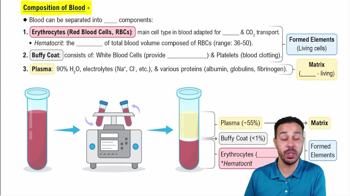
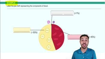
    Plasma and formed elements. Formed elements include erythrocytes, leukocytes, and platelets.
  • What is a buffy coat?

    The thin whitish layer of leukocytes and platelets between plasma and erythrocytes.
  • Definition of hematocrit

    The percentage of whole blood made up of red blood cells (RBCs). Male: 47% ± 5%. Female: 42% ± 5%.
  • Important characteristics/components of plasma

    pH 7.35–7.45. Albumin maintains osmotic pressure and transports substances. Gamma globulins are antibodies. Fibrinogen is involved in clotting.
  • Unusual features of mature erythrocytes

    No nucleus, essentially no organelles, and a biconcave shape.
  • Protein maintaining RBC biconcave shape

    Spectrin.
  • Normal RBC counts and lifespan

    Men: 5.1–5.8 × 10⁶ RBC/mm³. Women: 4.3–5.2 × 10⁶ RBC/mm³. Lifespan: 100–120 days. Diameter: 7.5 μm.
  • What is hemoglobin and its function?

    Hemoglobin binds and transports oxygen. It has 4 heme groups, 4 globin chains, and can carry 4 O₂ molecules.
  • Difference between hematopoiesis and erythropoiesis

    Hematopoiesis is production of any formed element. Erythropoiesis is production of erythrocytes only.
  • Sequence of erythropoiesis

    Hemocytoblast → proerythroblast → early erythroblast → late erythroblast → normoblast → reticulocyte → erythrocyte.
  • Nutrients necessary for erythropoiesis

    Nutrients, two B-complex vitamins, and iron.
  • How erythropoiesis is regulated

    ↓ O₂ → kidney cells hypoxic → kidneys release EPO → red bone marrow → ↑ RBC production → ↑ O₂.
  • Difference between transferrin, ferritin, and hemosiderin

    Transferrin transports iron in blood. Ferritin and hemosiderin store iron in cells.
  • How old erythrocytes are destroyed

    Macrophages destroy them in liver, spleen, and bone marrow. Hemoglobin separates into heme + globin.
  • What happens to hemoglobin when RBC is destroyed

    Globin → amino acids. Heme → iron + bilirubin. Iron reused/stored. Bilirubin travels to liver and enters bile.
  • Major RBC disorders

    Hemorrhagic anemia, hemolytic anemia, aplastic anemia, iron-deficiency anemia, pernicious anemia, thalassemia, sickle cell anemia, polycythemia.
  • How leukocytes differ from erythrocytes

    Leukocytes are larger, have nuclei, and function in immune and inflammatory responses.
  • Definitions of diapedesis, amoeboid motion, and positive chemotaxis

    Diapedesis: WBCs pass through vessel walls. Amoeboid motion: movement through tissues. Positive chemotaxis: movement toward chemical signals.
  • Granulocytes vs. agranulocytes

    Granulocytes: neutrophils, eosinophils, basophils. Agranulocytes: lymphocytes, monocytes.
  • Order of leukocytes from most to least abundant

    Neutrophils → lymphocytes → monocytes → eosinophils → basophils.
  • Main function of each leukocyte type

    Neutrophils phagocytize bacteria. Lymphocytes fight antigens and produce antibodies. Monocytes become macrophages. Eosinophils fight parasites and allergies. Basophils release histamine and heparin.
  • Definitions of leukocytosis, leukopenia, leukemia, infectious mononucleosis

    Leukocytosis: high WBC count. Leukopenia: low WBC count. Leukemia: malignant overproduction of leukocytes. Infectious mononucleosis involves B lymphocytes.
  • Origin and function of platelets

    Fragments of megakaryocytes. Normal count: 150,000–400,000. Main function: hemostasis.
  • What is hemostasis and its three major mechanisms

    Stoppage of bleeding. Mechanisms: vascular spasm, platelet plug formation, coagulation.
  • What happens during vascular spasm

    Injured vessel constricts. Serotonin and thromboxane A₂ enhance the spasm.
  • How a platelet plug forms

    Platelets stick to exposed collagen, activate, release chemicals, and recruit more platelets. ADP and thromboxane A₂ recruit more platelets.
  • Three phases of coagulation

    1. Formation of prothrombin activator. 2. Prothrombin → thrombin. 3. Fibrinogen → fibrin.
  • Difference between intrinsic and extrinsic clotting pathways

    Intrinsic: factors within blood. Extrinsic: requires tissue factor from outside blood.
  • Role of vitamin K in coagulation

    Required for synthesizing four clotting factors.
  • Clot retraction and fibrinolysis

    Clot retraction pulls wound edges closer. Fibrinolysis breaks down the clot during healing.
  • Definitions of thrombus, embolus, thrombocytopenia, hemophilia, DIC

    Thrombus: attached clot. Embolus: free-floating clot. Thrombocytopenia: low platelets. Hemophilia: clotting-factor defect. DIC: widespread clotting depleting clotting factors.
  • Difference between agglutinogen and agglutinin

    Agglutinogen: antigen on RBC surface. Agglutinin: antibody in plasma.
  • Antigens and antibodies associated with ABO blood types

    A: A antigen, anti-B antibody. B: B antigen, anti-A antibody. AB: A and B antigens, no anti-A or anti-B. O: no A or B antigens, anti-A and anti-B antibodies.
  • Why type O is universal donor and AB universal recipient

    O RBCs lack A and B antigens. AB blood has no anti-A or anti-B antibodies.
  • What is erythroblastosis fetalis

    Occurs when mother is Rh-negative and fetus Rh-positive, causing maternal antibodies to attack fetal RBCs.