Skip to main content
Anatomy & Physiology
나의 코스
배우기
시험 준비
AI 튜터
학습 가이드
교과서적 해결책
플래시카드
탐험해 보세요
앱을 사용해 보세요
나의 코스
배우기
시험 준비
AI 튜터
학습 가이드
교과서적 해결책
플래시카드
탐험해 보세요
앱을 사용해 보세요
뒤로
Spinal Cord and Nerve Anatomy - Anatomy & Physiology
카드를 뒤집기 위해 탭할 수 있습니다.
Length and width of the spinal cord
카드를 뒤집기 위해 탭할 수 있습니다.
👆
Length and width of the spinal cord
The spinal cord is about 18 inches (45 cm) long and 1/2 inch (14 mm) wide.
진행 상황 추적
컨트롤 버튼이 '내비게이션' 모드로 변경되었습니다.
1/25
추천 영상
0:50
Introduction to the Central Nervous System Example 1
12582
views
243
rank
2
comments
2:41
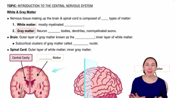
White & Gray Matter
12850
views
237
rank
3:34
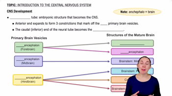
CNS Development
13500
views
269
rank
2
comments
이 집합의 용어 (25)
하이드의 정의
Length and width of the spinal cord
The spinal cord is about 18 inches (45 cm) long and 1/2 inch (14 mm) wide.
End of the spinal cord
The spinal cord ends between vertebrae L1 and L2 at the conus medullaris.
Number of spinal nerve pairs
There are 31 pairs of spinal nerves: 8 cervical, 12 thoracic, 5 lumbar, 5 sacral, and 1 coccygeal.
Spinal cord enlargements
Cervical enlargement supplies nerves to upper limbs; lumbar enlargement supplies nerves to pelvic region and lower limbs.
Cauda equina
Bundle of nerve roots from L2 to S5 occupying the vertebral canal below the conus medullaris.
Gray matter in spinal cord
Central butterfly-shaped area containing neuron cell bodies, with posterior (sensory) and anterior (motor) horns.
White matter in spinal cord
Surrounds gray matter; consists of myelinated axons organized into posterior, lateral, and anterior columns.
Function of gray commissure
Connects right and left sides of gray matter and contains the central canal filled with cerebrospinal fluid.
Ascending tracts
Carry sensory information from the body to the brain via first-, second-, and third-order neurons.
Gracile fasciculus
Carries signals for vibration, visceral pain, deep touch, and proprioception from lower limbs and trunk.
Cuneate fasciculus
Carries similar sensory signals as gracile fasciculus but from upper limbs and chest.
Spinothalamic tract
Carries pain, pressure, temperature, light touch, tickle, and itch sensations to the brain.
Spinoreticular tract
Transmits pain signals from tissue injury to the reticular formation and then to the cerebral cortex.
Spinocerebellar tracts
Carry proprioceptive signals from limbs and trunk to the cerebellum for coordination of movement.
Descending tracts
Carry motor commands from the brain to the spinal cord, involving upper and lower motor neurons.
Corticospinal tracts
Control precise, finely coordinated movements; most fibers decussate in the medulla forming lateral corticospinal tract.
Anatomy of a spinal nerve
Formed by the union of dorsal (sensory) and ventral (motor) roots; spinal nerves are mixed nerves.
Connective tissue layers of spinal nerves
Epineurium (outer), perineurium (around fascicles), and endoneurium (around individual axons).
Spinal meninges layers
Pia mater (inner), arachnoid mater (middle), and dura mater (outer) protect and isolate the spinal cord.
Function of muscle spindles
Stretch receptors in skeletal muscles that monitor muscle length and help coordinate movement and posture.
Monosynaptic reflex
Simplest reflex arc with sensory neuron synapsing directly on motor neuron, e.g., patellar tendon reflex.
Polysynaptic reflex
Reflex involving one or more interneurons between sensory and motor neurons, allowing complex responses.
Reciprocal inhibition
Inhibition of antagonistic muscles during a reflex to allow smooth movement, e.g., flexor reflex inhibits extensor muscles.
Crossed extensor reflex
Contralateral reflex that supports body weight by extending the opposite limb during a withdrawal reflex.
Dermatome
Bilateral region of skin monitored by a specific pair of spinal nerves; important for diagnosing nerve damage.
 뒤로뒤로
뒤로뒤로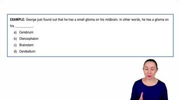
 0:50
0:50