Ch. 9 The Muscular System
Amerman - Human Anatomy & Physiology 2nd Edition
Amerman2nd EditionHuman Anatomy & PhysiologyISBN: 9780136873822당신이 사용하는 게 아니라요?교과서 변경
9장, 문제 9

Which of the erector spinae muscles is most lateral to the vertebral column?
a. Spinalis muscle
b. Iliocostalis muscle
c. Semispinalis muscle
d. Longissimus muscle

검증된 단계별 안내
1
Understand the anatomy of the erector spinae muscle group, which consists of three primary muscles: spinalis, longissimus, and iliocostalis. These muscles run parallel to the vertebral column and are responsible for extending and stabilizing the spine.
Identify the relative positions of the muscles within the erector spinae group. The spinalis muscle is the most medial, located closest to the vertebral column. The longissimus muscle is intermediate, positioned between the spinalis and iliocostalis muscles.
Recognize that the iliocostalis muscle is the most lateral of the erector spinae group. It is positioned furthest from the vertebral column and attaches to the ribs and iliac crest, contributing to lateral flexion and extension of the spine.
Exclude the semispinalis muscle from consideration, as it is not part of the erector spinae group. The semispinalis muscle belongs to the transversospinalis group and is located deeper in the back, closer to the vertebral column.
Conclude that the iliocostalis muscle is the correct answer because it is the most lateral muscle within the erector spinae group, based on its anatomical position and function.

비슷한 문제에 대한 검증된 영상 답변:

이 영상 해법은 위 문제에 도움이 된다고 튜터들이 추천한 것입니다.

주요 개념

질문에 올바르게 답하기 위해 반드시 이해해야 하는 핵심 개념들은 다음과 같습니다.

Erector Spinae Muscles

The erector spinae is a group of muscles that run along the spine and are responsible for extending and laterally flexing the vertebral column. This group consists of three primary muscles: iliocostalis, longissimus, and spinalis. Understanding their locations and functions is crucial for identifying which muscle is most lateral.
추천 영상:
2:00
Introduction to Muscles and Muscle Tissue Example 1

Muscle Anatomy and Positioning

In anatomy, the positioning of muscles relative to each other is essential for understanding their functions and interactions. The terms 'lateral' and 'medial' describe the position of structures in relation to the midline of the body. In the context of the erector spinae, knowing which muscle lies furthest from the vertebral column helps in answering questions about their anatomical arrangement.
추천 영상:
02:40
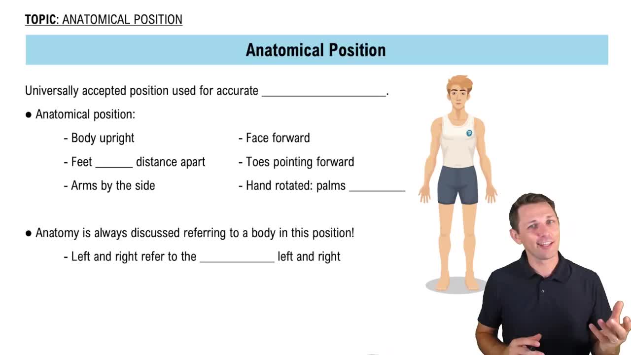
Anatomical Position

Spinal Muscle Functions

The erector spinae muscles play a vital role in maintaining posture and enabling movements of the spine, such as extension and rotation. Each muscle within this group has specific functions and contributes differently to spinal stability and mobility. Recognizing these functions aids in understanding why certain muscles are positioned as they are in relation to the vertebral column.
추천 영상:
가이드 코스
06:04
Functional Groups
관련 실천
교과서 질문

Mr. Bell presents with the inability to move certain muscles on one side of his face. You ask him to make various facial expressions and find that on his right side he is unable to purse his lips, pull in his cheeks, elevate his upper lip, and smirk. What muscles is Mr. Bell unable to contract?

686
views
교과서 질문

Which of the following muscle groups is considered muscles of inspiration?

a. External intercostal muscles

b. Internal intercostal muscles

c. External oblique muscles

d. Internal oblique muscles

855
views
교과서 질문

Match the muscle with its main action:

_____Sternocleidomastoid muscle     

_____Transversus abdominis muscle     

_____Internal oblique muscle     

_____Rectus abdominis muscle     

_____Splenius capitis muscle     

_____Quadratus lumborum muscle.

a. Laterally flexes the trunk

b. Flexes the trunk

c. Compresses the abdominal cavity

d. Extends the vertebral column

e. Flexes the head

f. Extends the head

711
views
교과서 질문

Which eye muscle passes through the trochlea and turns the eye inferiorly and laterally?

a. Lateral rectus muscle

b. Inferior rectus muscle

c. Inferior oblique muscle

d. Superior oblique muscle

1021
views
교과서 질문

Mark the following statements as true or false. If the statement is false, correct it to make a true statement.

The buccinator muscle pulls the cheeks in, producing sucking movements.

852
views
교과서 질문

The thick, bandlike muscle of mastication that covers much of the ramus of the mandible is the:

a. masseter muscle.

b. temporalis muscle.

c. lateral pterygoid muscle.

d. risorius muscle.

846
views