Ch. 15 The Special Senses
Amerman- Human Anatomy & Physiology 3e
Amerman3rd EditionHuman Anatomy & PhysiologyISBN: 9780138247201, 9780138247928, 9780138201814당신이 사용하는 게 아니라요?교과서 변경
15장, 문제 14

Explain how sounds of different frequencies are detected in the cochlea.

검증된 단계별 안내
1
Understand the structure of the cochlea: The cochlea is a spiral-shaped organ in the inner ear that contains the basilar membrane, which plays a key role in detecting sound frequencies. It is filled with fluid and lined with hair cells that respond to vibrations.
Learn about the basilar membrane's frequency mapping: The basilar membrane is tonotopically organized, meaning different regions of the membrane respond to different frequencies. High-frequency sounds are detected near the base of the cochlea (closer to the oval window), while low-frequency sounds are detected near the apex (the tip of the spiral).
Explore the role of hair cells: Hair cells are sensory receptors located on the basilar membrane. When sound waves cause the basilar membrane to vibrate, the hair cells in the corresponding region are stimulated. These cells convert mechanical vibrations into electrical signals.
Understand signal transmission: The electrical signals generated by the hair cells are transmitted to the auditory nerve. The auditory nerve carries these signals to the brain, where they are interpreted as specific frequencies of sound.
Connect the concept to frequency discrimination: The ability to detect different frequencies is due to the precise mechanical properties of the basilar membrane and the specific activation of hair cells in distinct regions. This allows the cochlea to act as a frequency analyzer, separating sounds into their component frequencies.

비슷한 문제에 대한 검증된 영상 답변:

이 영상 해법은 위 문제에 도움이 된다고 튜터들이 추천한 것입니다.

주요 개념

질문에 올바르게 답하기 위해 반드시 이해해야 하는 핵심 개념들은 다음과 같습니다.

Cochlea Structure

The cochlea is a spiral-shaped, fluid-filled structure in the inner ear that plays a crucial role in hearing. It contains the basilar membrane, which varies in width and stiffness along its length, allowing it to respond differently to various sound frequencies. High-frequency sounds stimulate the base of the cochlea, while low-frequency sounds affect the apex, enabling the detection of a wide range of pitches.
추천 영상:
4:16
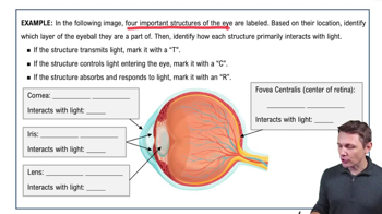
Structure of the Eyeball Example 1

Frequency Encoding

Frequency encoding in the cochlea refers to the process by which different sound frequencies are translated into neural signals. This is achieved through a mechanism called tonotopic organization, where specific areas of the basilar membrane correspond to specific frequencies. As sound waves travel through the cochlear fluid, they cause the membrane to vibrate, activating hair cells that convert these vibrations into electrical impulses sent to the brain.
추천 영상:
01:10
The Refractory Period Example 1

Hair Cells and Signal Transduction

Hair cells are the sensory receptors located on the basilar membrane of the cochlea. When sound vibrations cause the basilar membrane to move, the hair cells bend, leading to the opening of ion channels and the generation of electrical signals. These signals are then transmitted via the auditory nerve to the brain, where they are interpreted as sound, allowing us to perceive different frequencies.
추천 영상:
가이드 코스
07:02
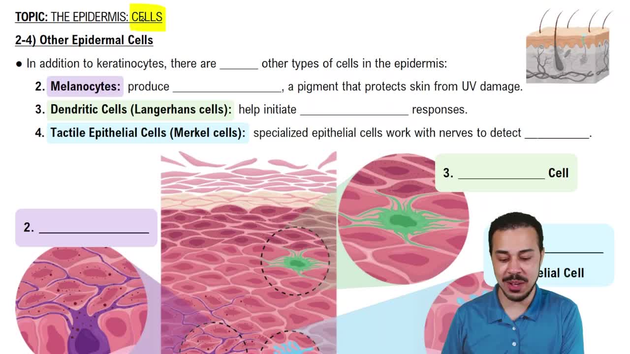
Other Epidermal Cells