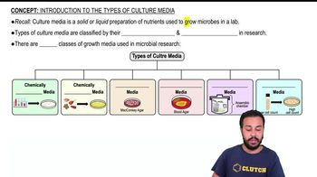
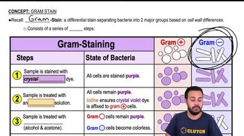
A bacterial specimen exhibits the following growth on blood agar. What can you most reasonably conclude about the bacterium? Select all that apply. a. It is not S. pyogenes. b. It is alpha hemolytic. c. It is beta hemolytic. d. It is gamma hemolytic. e. It is Gram-positive. <Image>
검증된 단계별 안내
1
Step 1: Understand the types of hemolysis on blood agar. Blood agar is used to differentiate bacteria based on their ability to lyse red blood cells, producing three types of hemolysis: alpha (partial hemolysis, greenish discoloration), beta (complete hemolysis, clear zone), and gamma (no hemolysis, no change).
Step 2: Identify the hemolysis pattern shown by the bacterial specimen on the blood agar plate. This is crucial because it helps narrow down the bacterial species or group.
Step 3: Recall that Streptococcus pyogenes is typically beta hemolytic, meaning it produces a clear zone of complete hemolysis around colonies on blood agar.
Step 4: Use the hemolysis pattern observed to determine which options are reasonable. For example, if the specimen shows beta hemolysis, then it could be S. pyogenes and is beta hemolytic; if alpha hemolysis is observed, it is not S. pyogenes but could be other Streptococcus species like S. pneumoniae.
Step 5: Consider Gram staining characteristics. Most hemolytic streptococci are Gram-positive cocci, so if the specimen is a streptococcus, it is likely Gram-positive. This helps decide if option (e) is reasonable.
비슷한 문제에 대한 검증된 영상 답변:
이 영상 해법은 위 문제에 도움이 된다고 튜터들이 추천한 것입니다.
영상 길이:
3m
영상 재생:
0 댓글
주요 개념
질문에 올바르게 답하기 위해 반드시 이해해야 하는 핵심 개념들은 다음과 같습니다.
Hemolysis Types on Blood Agar
Hemolysis refers to the breakdown of red blood cells around bacterial colonies on blood agar. Alpha hemolysis causes partial, greenish discoloration; beta hemolysis results in clear, complete lysis; gamma hemolysis indicates no hemolysis or change. Identifying hemolysis type helps differentiate bacterial species.
Streptococcus pyogenes is a Gram-positive bacterium known for beta hemolysis on blood agar, producing clear zones around colonies. Recognizing its hemolytic pattern is key to distinguishing it from other streptococci and related bacteria.
Gram staining differentiates bacteria into Gram-positive (purple) and Gram-negative (pink) based on cell wall structure. This classification aids in identifying bacteria and predicting their behavior, including antibiotic susceptibility and pathogenicity.