Skip to main content
Microbiology
나의 코스
배우기
시험 준비
AI 튜터
학습 가이드
교과서적 해결책
플래시카드
탐험해 보세요
앱을 사용해 보세요
나의 코스
배우기
시험 준비
AI 튜터
학습 가이드
교과서적 해결책
플래시카드
탐험해 보세요
앱을 사용해 보세요
뒤로
Light Microscopes that Detect Fluorescence quiz
카드를 뒤집기 위해 탭할 수 있습니다.
What is fluorescence in the context of microscopy?
카드를 뒤집기 위해 탭할 수 있습니다.
👆
What is fluorescence in the context of microscopy?
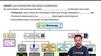
Fluorescence is the ability to absorb short wavelengths of light and immediately emit longer wavelengths of visible light.
진행 상황 추적
컨트롤 버튼이 '내비게이션' 모드로 변경되었습니다.
1/15
관련 플래시카드
관련 실천
추천 영상
Light Microscopes that Detect Fluorescence definitions
Light Microscopes that Detect Fluorescence
13 용어
Light Microscopes that Detect Fluorescence
9. Microscopes
5 문제점
주제
Monica
Electron Microscopes
9. Microscopes
3 문제점
주제
Brendan
9. Microscopes - Part 1 of 3
6 주제
12 문제점
장
Nicole
9. Microscopes - Part 2 of 3
8 주제
10 문제점
장
Nicole
9. Microscopes - Part 3 of 3
1 주제
3 문제점
장
01:46
Light Microscopes that Detect Fluorescence
2075
views
40
rank
1
comments
02:54
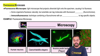
Fluorescence Microscopes
1956
views
32
rank
02:01
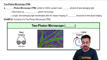
Two-Photon Microscope (TPM)
1781
views
32
rank
이 집합의 용어 (15)
하이드의 정의
What is fluorescence in the context of microscopy?
Fluorescence is the ability to absorb short wavelengths of light and immediately emit longer wavelengths of visible light.
How do fluorescent molecules appear under a fluorescence microscope?
Fluorescent molecules appear as bright objects against a darker background.
What type of light do fluorescence microscopes use to illuminate specimens?
Fluorescence microscopes use ultraviolet light to cause specimens to fluoresce.
What is the purpose of tagging molecules with fluorescent dyes in microscopy?
Tagging molecules with fluorescent dyes allows specific molecules to fluoresce, making them visible and trackable under the microscope.
What is immunofluorescence?
Immunofluorescence is a technique that combines fluorescent dyes with antibodies to tag and visualize specific molecules within cells.
What advantage does fluorescence microscopy provide in imaging?
Fluorescence microscopy creates vibrant and colorful images, allowing for the visualization of specific structures or molecules.
What is a confocal scanning laser microscope (CSLM) and what is its main feature?
A CSLM is a computer-controlled microscope that couples a laser to a fluorescence microscope, generating high-contrast, 3D images of specimens.
How does a CSLM allow for examination of specimens?
A CSLM allows the viewer to access several different planes of focus inside the specimen, enabling detailed examination of different layers.
What is a two-photon microscope (TPM) and how does it differ from a CSLM?
A TPM uses two photons and longer, less damaging wavelengths of light, allowing for deeper imaging of thick structures compared to CSLMs.
What unique feature does two-photon microscopy offer?
Two-photon microscopy allows for time-lapse imaging, creating video-like effects of dynamic processes within thick specimens.
What is the main advantage of super resolution microscopes (SRMs) over traditional light microscopes?
SRMs provide a much higher resolution of about 0.01 micrometers, allowing for the distinction of objects that are extremely close together.
How does the resolution of SRMs compare to pre-2014 light microscopes?
SRMs have about 20 times better resolution than traditional light microscopes, which had a maximum resolution of 0.2 micrometers.
What can SRMs allow scientists to visualize that was not possible before?
SRMs can allow scientists to visualize and track even single molecules within cells due to their extremely high resolution.
Why are super resolution microscopes compared to HDTVs in the lesson?
SRMs are compared to HDTVs because they provide much clearer and more detailed images, similar to the difference between an old TV and a modern HDTV.
What is the significance of using less damaging light in two-photon microscopy?
Using less damaging light in two-photon microscopy reduces harm to specimens and enables deeper imaging of thick biological structures.
 뒤로뒤로
뒤로뒤로
 01:46
01:46