Skip to main content
Microbiology
나의 코스
배우기
시험 준비
AI 튜터
학습 가이드
교과서적 해결책
플래시카드
탐험해 보세요
앱을 사용해 보세요
나의 코스
배우기
시험 준비
AI 튜터
학습 가이드
교과서적 해결책
플래시카드
탐험해 보세요
앱을 사용해 보세요
뒤로
Reviewing the Different Types of Microscopes definitions
카드를 뒤집기 위해 탭할 수 있습니다.
Bright Field Microscope
카드를 뒤집기 위해 탭할 수 있습니다.
👆
Bright Field Microscope
Most common light microscope; observes stained or unstained specimens on a bright background, but may have poor contrast for transparent cells.
진행 상황 추적
컨트롤 버튼이 '내비게이션' 모드로 변경되었습니다.
1/14
관련 플래시카드
관련 실천
추천 영상
Reviewing the Different Types of Microscopes quiz
Reviewing the Different Types of Microscopes
15 용어
Reviewing the Different Types of Microscopes
9. Microscopes
4 문제점
주제
Nicole
Introduction to Staining
9. Microscopes
주제
9. Microscopes - Part 1 of 3
6 주제
12 문제점
장
Nicole
9. Microscopes - Part 2 of 3
8 주제
10 문제점
장
Nicole
9. Microscopes - Part 3 of 3
1 주제
3 문제점
장
05:32
Reviewing the Different Types of Microscopes
2162
views
28
rank
이 집합의 용어 (14)
하이드의 정의
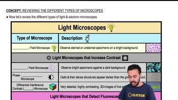
Bright Field Microscope
Most common light microscope; observes stained or unstained specimens on a bright background, but may have poor contrast for transparent cells.
Compound Light Microscope
A type of bright field microscope with multiple lenses, allowing calculation of total magnification for specimen observation.
Dark Field Microscope
Produces a dark background, making specimens appear brighter and enhancing contrast for better visualization.
Phase Contrast Microscope
Enhances visibility of dense cell structures, making them appear darker against a grayish background without staining.
Differential Interference Contrast Microscope
Generates highly detailed, three-dimensional images of live specimens with enhanced contrast.
Confocal Scanning Laser Microscope
Provides high contrast and three-dimensional imaging by focusing on multiple planes within a specimen.
Two-Photon Microscope
Enables high contrast, three-dimensional, and time-lapse imaging of deep structures, similar to x-ray vision.
Super Resolution Microscope
Offers extremely high resolution, around 0.01 micrometers, surpassing standard light microscopes.
Electron Microscope
Uses electrons instead of visible light to magnify specimens, allowing for much higher resolution imaging.
Transmission Electron Microscope
Creates two-dimensional images by passing electrons through specimens, ideal for visualizing internal cell structures.
Scanning Electron Microscope
Generates three-dimensional images by detecting electrons scattered from specimen surfaces, revealing external structures.
Resolution
The ability to distinguish fine details and structures, with higher values allowing clearer, more detailed images.
Contrast
The difference in visual properties that makes an object distinguishable from its background, crucial for specimen visualization.
Magnification
The process of enlarging the appearance of a specimen, allowing for detailed observation of microscopic structures.
 뒤로뒤로
뒤로뒤로
 05:32
05:32