Ch. 3 - Observing Microorganisms Through a Microscope
Tortora - Microbiology: An Introduction 14th Edition
Tortora14th EditionMicrobiology: An IntroductionISBN: 9780138200398당신이 사용하는 게 아니라요?교과서 변경
3장, 문제 3

Carbolfuchsin can be used as a simple stain and a negative stain. As a simple stain, the pH is:
a. 2
b. Higher than the negative stain
c. Lower than the negative stain
d. The same as the negative stain

검증된 단계별 안내
1
Understand the role of carbolfuchsin in staining: it can be used both as a simple stain and as a negative stain, but the pH conditions differ depending on the staining method.
Recall that a simple stain typically uses a basic dye, which means the pH is usually higher (alkaline) to allow the dye to bind to negatively charged cell components.
Recognize that a negative stain uses an acidic dye, which means the pH is lower (acidic) so that the background is stained and the cells remain unstained.
Compare the pH of carbolfuchsin when used as a simple stain (basic pH) versus when used as a negative stain (acidic pH).
Conclude that the pH of carbolfuchsin as a simple stain is higher than the pH when it is used as a negative stain.

비슷한 문제에 대한 검증된 영상 답변:

이 영상 해법은 위 문제에 도움이 된다고 튜터들이 추천한 것입니다.
영상 길이:
2m
도움이 되었나요?

주요 개념

질문에 올바르게 답하기 위해 반드시 이해해야 하는 핵심 개념들은 다음과 같습니다.

Carbolfuchsin as a Stain

Carbolfuchsin is a basic dye commonly used in microbiology for staining bacterial cells. It binds to negatively charged components like nucleic acids and cell walls, making cells visible under a microscope. Its staining properties depend on the pH of the solution, which affects its charge and interaction with cells.
추천 영상:
가이드 코스
02:10
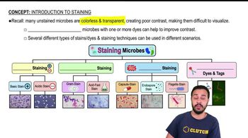
Introduction to Staining

Simple Stain vs. Negative Stain

A simple stain colors the cells directly, usually with a basic dye, highlighting cell morphology. In contrast, a negative stain colors the background, leaving cells unstained and visible as clear shapes. The pH of the stain influences whether the dye acts as a basic or acidic stain, affecting which part is stained.
추천 영상:
가이드 코스
03:23
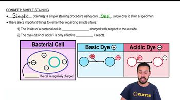
Simple Staining

Effect of pH on Dye Charge and Staining

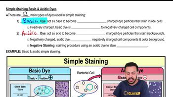
The pH of a staining solution determines the ionization state of the dye molecules. At low pH, dyes like carbolfuchsin carry a positive charge, acting as basic stains that bind to negatively charged cell components. At higher pH, the dye may lose its positive charge, altering its staining behavior, such as in negative staining.
추천 영상:
가이드 코스
05:30
Simple Staining Basic & Acidic Dyes
관련 실천
교과서 질문

Assume you stain Bacillus by applying malachite green with heat and then counterstain with safranin. Through the microscope, the green structures are

a. Cell walls

b. Capsules

c. Endospores

d. Flagella

e. Impossible to identify

1899
views
교과서 질문

Three-dimensional images of live cells can be produced with:

a. Darkfield microscopy

b. Fluorescence microscopy

c. Transmission electron microscopy

d. Confocal microscopy

e. Phase-contrast microscopy

1659
views
교과서 질문

The maximum magnification of a compound microscope is (a) ________; that of an electron microscope, (b) ________. The maximum resolution of a compound microscope is (c) ________; that of an electron microscope, (d) ________. One advantage of a scanning electron microscope over a transmission electron microscope is (e) ________.

1905
views
교과서 질문

Looking at the cell of a photosynthetic microorganism, you observe the chloroplasts are green in brightfield microscopy and red in fluorescence microscopy. You conclude:

a. Chlorophyll is fluorescent

b. The magnification has distorted the image

c. You’re not looking at the same structure in both microscopes

d. The stain masked the green color

e. None of the above

1179
views
교과서 질문

Calculate the total magnification of the nucleus of a cell being observed through a compound light microscope with a 10x ocular lens and an oil immersion lens.

1828
views
교과서 질문

Which type of microscope would be best to use to observe each of the following?

a. A stained bacterial smear

b. Unstained bacterial cells: the cells are small, and no detail is needed

c. Unstained live tissue when it is desirable to see some intracellular detail

d. A sample that emits light when illuminated with ultraviolet light

e. Intracellular detail of a cell that is 1μm long

f. Unstained live cells in which intracellular structures are shown in color

1799
views