Organization of the Body: Serous Membrane Locations definitions Flashcards
 Back
BackOrganization of the Body: Serous Membrane Locations definitions
You can tap to flip the card.
Control buttons has been changed to "navigation" mode.
1/15
Serous Membranes
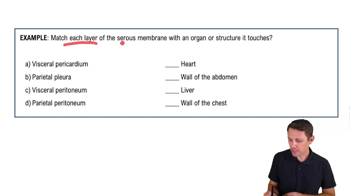
Thin tissues that line certain internal cavities, reducing friction between organs during movement.Pleurae
Membranes forming pleural cavities, surrounding each lung, allowing lung movement without friction.Pericardium
Membrane forming the pericardial cavity, surrounding the heart, facilitating its movement.Peritoneum
Membrane forming the peritoneal cavity, surrounding most digestive organs, allowing complex organ movement.Visceral Layer
The part of a serous membrane that directly contacts and covers an organ.Parietal Layer
The part of a serous membrane that lines the body wall or cavity.Pleural Cavities
Spaces created by pleurae surrounding each lung, allowing lung expansion and contraction.Pericardial Cavity
Space formed by the pericardium surrounding the heart, enabling its movement.Peritoneal Cavity
Complex space formed by the peritoneum, surrounding various digestive organs.Visceral Pericardium
The layer of the pericardium that directly touches the heart.Parietal Pleura
The layer of pleura that lines the chest wall, surrounding the lungs.Visceral Peritoneum
The layer of the peritoneum that directly contacts abdominal organs.Parietal Peritoneum
The layer of the peritoneum that lines the abdominal wall.Thoracic Cavity
The body cavity housing the lungs and heart, enclosed by the rib cage.Abdominal Cavity
The body cavity containing most digestive organs, part of the peritoneal cavity.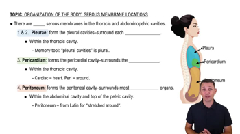
 3:55
3:55