Support, protection, anchorage for muscles, mineral storage (calcium & phosphate), blood cell formation (hematopoiesis), and triglyceride storage in yellow marrow.
Amorphous but firm matrix with collagen fibers; chondrocytes in lacunae; supports and reinforces; found in embryonic skeleton, articular cartilage, costal cartilage, nose, trachea, and larynx.
Elastic cartilage characteristics
Similar to hyaline but with more elastic fibers; maintains shape with flexibility; located in external ear (pinna) and epiglottis.
Fibrocartilage characteristics
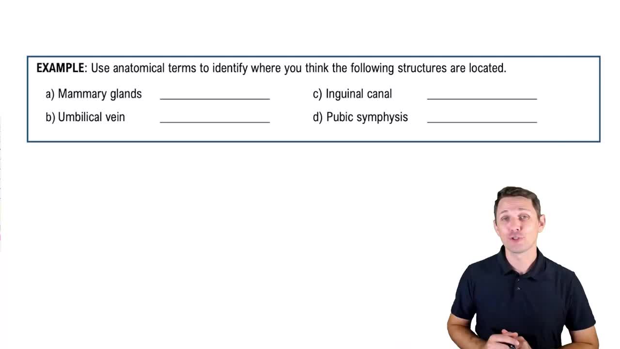
Matrix less firm than hyaline with thick collagen fibers; tensile strength absorbs compressive shock; found in intervertebral discs, pubic symphysis, and knee menisci.
Classification of bones by shape
Long bones (longer than wide), short bones (cube-shaped), flat bones (thin, flattened), and irregular bones (complex shapes).
Long bone anatomy: diaphysis
Tubular shaft forming the long axis; thick collar of compact bone surrounding the medullary cavity filled with yellow bone marrow in adults.
Long bone anatomy: epiphyses
Bone ends with outer compact bone and inner spongy bone; covered with articular (hyaline) cartilage; separated from diaphysis by epiphyseal line.
Periosteum layers
Outer fibrous layer of dense irregular connective tissue; inner osteogenic layer containing osteoprogenitor cells, osteoblasts, and osteoclasts.
Endosteum
Membrane lining internal bone surfaces including trabeculae and canals; contains osteoprogenitor cells, osteoblasts, and osteoclasts.
Bone marrow types and locations
Red marrow in infants fills medullary cavity and spongy bone; in adults, red marrow is in trabecular cavities of spongy bone in proximal epiphyses and diploë of flat bones; yellow marrow stores fat.
Structural unit of compact bone; concentric lamellae of bone matrix with collagen fibers alternating directions; central canal contains blood vessels and nerves.
Canals in compact bone
Central canal runs through osteon center; perforating canals connect central canals to medullary cavity; lacunae house osteocytes connected by canaliculi.
Spongy bone structure
Contains trabeculae aligned with stress lines; irregular lamellae and osteocytes connected by canaliculi; lacks osteons.
Chemical composition of bone
Organic components: bone cells and osteoid (ground substance and collagen fibers); inorganic components: mineral salts, mainly hydroxyapatite (65%).
Endochondral ossification
Bone develops by replacing hyaline cartilage; forms most bones below the skull except clavicles.
Intramembranous ossification
Bone develops from fibrous connective tissue membranes; forms flat bones of the skull and clavicles.
Zones of long bone growth in length
Proliferation zone, hypertrophic zone, calcification zone, and ossification zone in the epiphyseal plate.
Appositional growth
Bone growth in thickness; osteoblasts add bone matrix on external surface, osteoclasts remove bone on endosteal surface.
Bone remodeling
Continuous process of bone resorption by osteoclasts and deposition by osteoblasts; about 5-10% of bone replaced yearly.
Role of osteoclasts in bone resorption
Multinucleated cells that break down bone matrix by secreting acid and enzymes; create grooves and release minerals into bloodstream.
Role of osteoblasts in bone deposition
Secrete osteoid which calcifies; proteins bind calcium triggering mineralization and formation of hydroxyapatite crystals.
Hormones regulating calcium homeostasis
Parathyroid hormone (PTH) and calcitriol regulate blood calcium levels via bone remodeling.
Wolff's law
Bone grows or remodels in response to mechanical stress and gravitational forces.
Fracture classifications by bone end position
Non-displaced (bone ends retain normal position) and displaced (bone ends out of normal alignment).
Fracture classifications by completeness
Complete (bone broken through) and incomplete (partial break).
Fracture classifications by skin penetration
Open (compound) fractures penetrate skin; closed (simple) fractures do not.
 Back
 02:52