 Back
BackAnatomy & Physiology: Bones, Joints, and Skeletal Tissues
 01:27
01:27
Terms in this set (25)
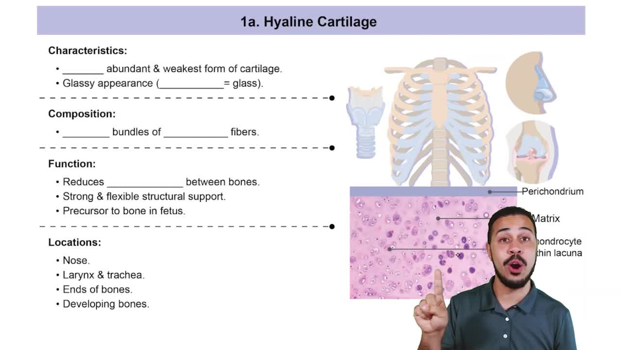
Hyaline cartilage: support, flexibility, resilience; most abundant.
Elastic cartilage: similar to hyaline but with elastic fibers; found in external ear and epiglottis.
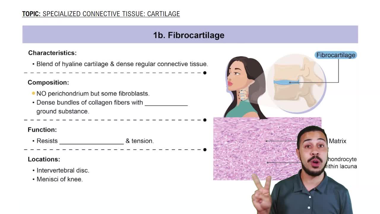
Fibrocartilage: highly compressed with tensile strength; found in menisci and intervertebral discs.
Red bone marrow: in infants, found in medullary cavity and all spongy bone areas; in adults, found in diploë of flat bones and heads of femur and humerus.
Yellow bone marrow: found in medullary cavity of long bones, mainly fat.
Consists of a diaphysis (shaft) made of compact bone surrounding the medullary cavity with yellow marrow, and epiphyses (ends) with compact bone exterior and spongy bone interior, covered by articular cartilage.
Support the body framework, protect vital organs, enable movement via muscle attachment, store minerals (calcium, phosphorus), and produce blood cells (hematopoiesis).
Projections: sites for muscle/ligament attachment (e.g., tuberosity, crest).
Joint surfaces: form joints (e.g., head, facet).
Depressions and openings: allow passage of vessels/nerves (e.g., foramen, fossa).
The structural unit of compact bone, consisting of concentric lamellae around a central (Haversian) canal containing blood vessels and nerves.
Organic: osteoid (collagen and proteins) provides flexibility and tensile strength.
Inorganic: hydroxyapatites (calcium phosphate salts) provide hardness and resistance to compression.
Bone develops from fibrous connective tissue membranes; forms flat bones like skull and clavicles; involves ossification center formation, matrix secretion, woven bone and periosteum formation, then bone collar and red marrow development.
Bone forms by replacing hyaline cartilage; involves bone collar formation, cartilage cavitation, periosteal bud invasion, spongy bone formation, medullary cavity development, and secondary ossification centers in epiphyses.
Growth zone: cartilage cells undergo mitosis.
Transformation zone: cells enlarge, matrix calcifies, cells die.
Osteogenic zone: new bone formation occurs.
Growth hormone stimulates epiphyseal plate activity in infancy and childhood; testosterone and estrogens promote adolescent growth spurts and later induce epiphyseal plate closure, ending longitudinal growth.
A bone grows or remodels in response to the mechanical forces or demands placed upon it, leading to changes in bone thickness, trabeculae alignment, and projections where muscles attach.
Position: nondisplaced or displaced.
Completeness: complete or incomplete.
Orientation: linear or transverse.
Skin penetration: compound (open) or simple (closed).
1. Hematoma formation.
2. Fibrocartilaginous callus formation.
3. Bony callus formation.
4. Bone remodeling.
Both caused by insufficient calcium or vitamin D deficiency.
Osteomalacia: soft, weakened bones in adults with pain on weight bearing.
Rickets: similar in children, causing bowed legs and bone deformities.
A disease where bone resorption outpaces deposition, leading to fragile bones; most common in postmenopausal women.
Based on material binding bones and presence of joint cavity: fibrous (no cavity), cartilaginous (no cavity), and synovial (fluid-filled cavity).
Synarthroses: immovable.
Amphiarthroses: slightly movable.
Diarthroses: freely movable.
Sutures: skull bones.
Syndesmoses: tibia-fibula connection.
Gomphoses: tooth in alveolar socket.
Synchondroses: hyaline cartilage, immovable (e.g., epiphyseal plates).
Symphyses: fibrocartilage pad, slightly movable (e.g., intervertebral discs, pubic symphysis).
Articular cartilage, joint cavity, articular capsule, synovial fluid, and reinforcing ligaments.
Articular surface shape, ligament number and location, and muscle tone via tendons crossing the joint.
Plane: flat surfaces, gliding (intercarpal).
Hinge: uniaxial flexion/extension (elbow).
Pivot: uniaxial rotation (proximal radioulnar).
Condyloid: biaxial angular motion (wrist).
Saddle: biaxial, greater movement (thumb carpometacarpal).
Ball-and-socket: multiaxial (shoulder, hip).
Sprains: stretched or torn ligaments.
Cartilage injuries: torn cartilage, often repaired arthroscopically.
Dislocations: bones forced out of alignment, often with sprains and inflammation.
Osteoarthritis: wear-and-tear, cartilage breakdown, common with aging.
Rheumatoid arthritis: autoimmune, chronic inflammation, pannus formation, joint deformity.