Skip to main content
Anatomy & Physiology
My Course
Learn
Exam Prep
AI Tutor
Study Guides
Textbook Solutions
Flashcards
Explore
Try the app
My Course
Learn
Exam Prep
AI Tutor
Study Guides
Textbook Solutions
Flashcards
Explore
Try the app
Back
Anatomy & Physiology: Connective Tissue, Cartilage, Bone, Muscle, Skin, and Upper Limb Anatomy
1 student found this helpful
You can tap to flip the card.
What do all connective tissues have in common?
You can tap to flip the card.
👆
What do all connective tissues have in common?
Cells are surrounded by an
extracellular matrix (ECM)
made of protein fibers and ground substance.
Track progress
Control buttons has been changed to "navigation" mode.
1/92
Recommended videos
Guided course
06:37
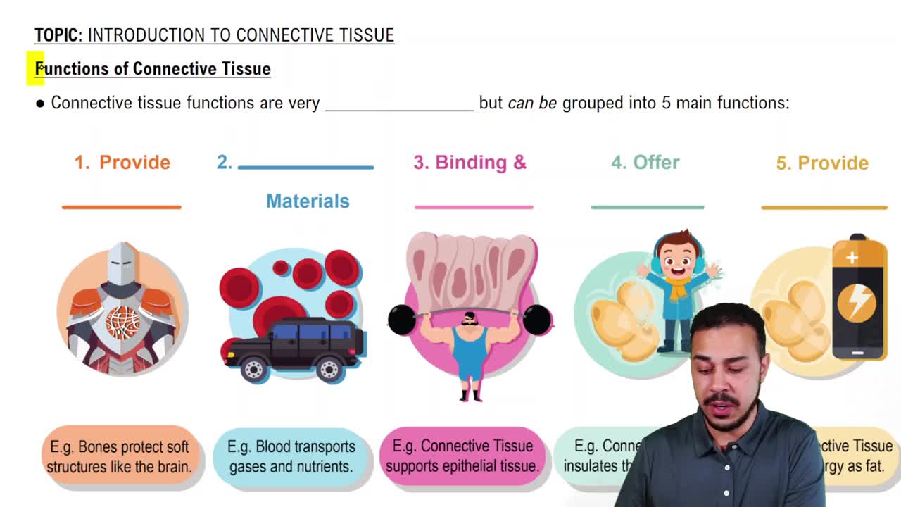
Functions of Connective Tissue
8177
views
137
rank
Guided course
08:54
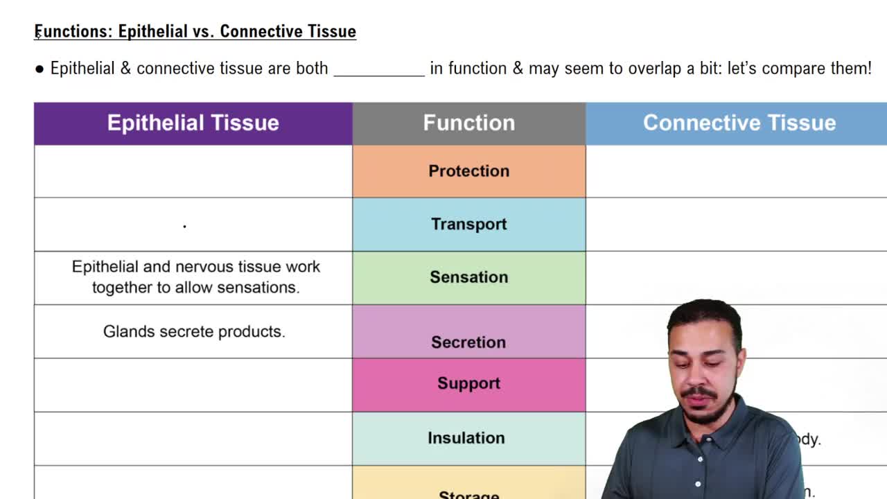
Functions: Epithelial vs. Connective Tissue
7722
views
120
rank
3
comments
Guided course
01:32
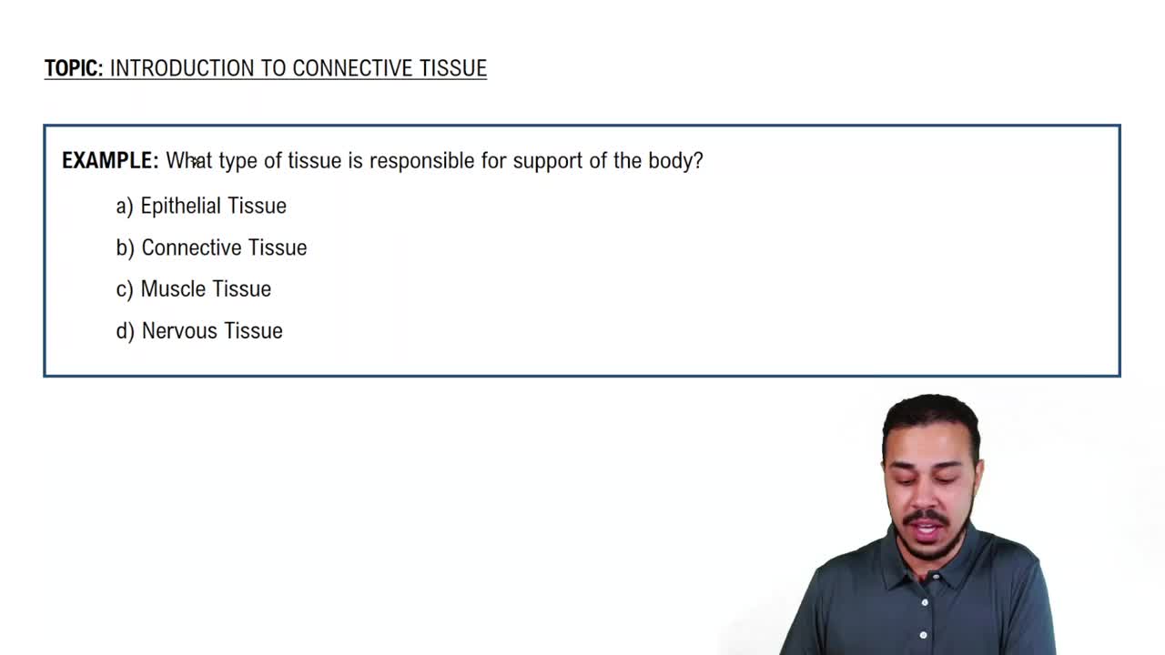
Introduction to Connective Tissue Example 1
6469
views
65
rank
1
comments
Terms in this set (92)
Hide definitions
What do all connective tissues have in common?
Cells are surrounded by an
extracellular matrix (ECM)
made of protein fibers and ground substance.
Functions of connective tissue
Bind and support tissues; protect organs; store energy; insulate; transport substances; immune defense; tissue repair.
Where is connective tissue proper found?
Throughout the body, especially under epithelia, around organs, between tissues, and in tendons and ligaments.
Cells found in connective tissue proper
Adipocytes, mast cells, macrophages, and fibroblasts.
What are adipocytes?
Fat cells that store lipids.
Role of mast cells
Immune cells involved in inflammation.
Role of macrophages
Ingest foreign substances and cellular debris through phagocytosis.
Role of fibroblasts
Produce protein fibers and ground substance of the ECM.
Primary component of loose connective tissue
Ground substance.
Main function of loose connective tissue
Wraps, cushions, supports, and binds tissues.
Primary component of dense connective tissue
Protein fibers, especially collagen fibers.
Types of dense connective tissue
Dense irregular, dense regular, and elastic connective tissue.
Collagen fiber arrangement in dense irregular connective tissue
Randomly/irregularly arranged to resist tension from many directions.
Location of dense irregular connective tissue
Dermis of the skin and capsules around organs and joints.
Collagen fiber arrangement in dense regular connective tissue
Parallel to each other to resist strong pulling forces in one direction.
Location of dense regular connective tissue
Tendons, ligaments, and aponeuroses.
Primary component of elastic connective tissue
Elastic fibers.
Location of elastic connective tissue
Walls of large arteries, some ligaments, and vocal ligaments.
What is reticular tissue?
A network of thin reticular fibers forming a supportive framework.
Location of reticular tissue
Lymph nodes, spleen, and bone marrow.
Functions of adipose tissue
Stores energy, provides insulation, cushions/protects organs, and helps regulate metabolism.
Key properties of cartilage
Strong but flexible, supportive, avascular, and slow to heal.
Cell producing most ECM in cartilage
Chondroblast.
Mature cartilage cell
Chondrocyte located in lacunae.
Why is cartilage slow to heal?
Because it is avascular and receives nutrients by diffusion through the ECM.
Three types of cartilage
Hyaline, fibrocartilage, and elastic cartilage.
Most abundant cartilage type
Hyaline cartilage.
ECM dominance in hyaline cartilage
Collagen fibers, especially type II collagen.
Locations of hyaline cartilage
Articular surfaces, trachea, nose, costal cartilage, larynx, and fetal skeleton.
ECM dominance in fibrocartilage
Thick collagen fibers, especially type I collagen.
Locations of fibrocartilage
Intervertebral discs, pubic symphysis, and knee menisci.
Difference of elastic cartilage
Contains many elastic fibers, making it more flexible.
Locations of elastic cartilage
External ear and epiglottis.
Bone tissue other name
Osseous tissue.
Bone-building cell
Osteoblast.
Mature bone cell
Osteocyte located in lacunae.
Bone-resorbing cell
Osteoclast.
Unique feature of blood as connective tissue
Its extracellular matrix is fluid, called plasma.
Function of red blood cells
Transport oxygen and carbon dioxide.
Function of white blood cells
Immune defense.
What is a platelet?
A cell fragment involved in blood clotting.
Unique property of muscle tissue
Specialized for contraction and movement.
Muscle type that is striated and voluntary
Skeletal muscle.
Muscle type that is non-striated and involuntary
Smooth muscle.
Muscle type that is striated and involuntary
Cardiac muscle.
Location of skeletal muscle
Attached to bones.
Appearance of skeletal muscle
Long, cylindrical, striated, and multinucleated fibers.
Location of smooth muscle
Walls of organs and blood vessels.
Appearance of smooth muscle
Spindle-shaped cells with one nucleus and no striations.
Location of cardiac muscle
Heart.
Appearance of cardiac muscle
Branched, striated cells with one nucleus and intercalated discs.
Where is nervous tissue found?
Brain, spinal cord, and peripheral nerves.
Two main types of nervous tissue cells
Neurons and neuroglial cells (glia).
Main function of neurons
Receive and transmit electrical signals.
Main function of neuroglial cells
Support, protect, nourish, and maintain neurons.
Largest organ in the body
Skin (cutaneous membrane).
Two components of skin
Epidermis and dermis.
What is the epidermis?
The superficial/outer layer of skin made of keratinized stratified squamous epithelium.
Does the epidermis contain blood vessels?
No, it is avascular and receives nutrients by diffusion from the dermis.
What is the dermis?
The deeper connective tissue layer beneath the epidermis.
Accessory structures of the skin
Hair, nails, sweat glands, sebaceous glands, and sensory receptors.
Functions of skin
Protection, sensation, thermoregulation, vitamin D production, prevention of water loss, excretion, and immune defense.
What is the acid mantle?
A slightly acidic layer on the skin surface that inhibits microorganism growth.
How does skin create a waterproof barrier?
Keratin and lipids form a water-resistant barrier preventing excessive water loss.
Why is sensation important to homeostasis?
It allows detection of temperature, pain, pressure, and other changes to respond appropriately.
What is thermoregulation?
The process of maintaining a stable body temperature.
Body response when too hot
Sweating and vasodilation increase heat loss.
Body response when too cold
Vasoconstriction conserves heat and shivering produces heat.
Why is vitamin D important?
It helps with calcium absorption and bone health.
Most common cell in the epidermis
Keratinocyte.
What do keratinocytes produce?
Keratin, which makes skin strong, protective, and water-resistant.
What junction connects keratinocytes?
Desmosomes, which help resist mechanical stress.
Five epidermal layers from superficial to deep
Stratum corneum, stratum lucidum, stratum granulosum, stratum spinosum, stratum basale.
Mnemonic for epidermal layers
Come Let's Get Sun Burned.
Deepest epidermal layer
Stratum basale, one cell layer thick with mostly cuboidal to columnar cells.
Function of stratum basale
Produces new keratinocytes via rapid cell division.
What does UV radiation allow skin to produce?
A vitamin D precursor that can become vitamin D.
Why is the stratum spinosum called the spiny layer?
Cells appear spiny due to desmosomes under a microscope.
Function of stratum granulosum
Contains granules that release lipids to create a water-resistant barrier.
Where is the stratum lucidum found?
Only in thick skin, such as palms and soles.
Most superficial epidermal layer
Stratum corneum, composed of dead, keratinized cells that shed.
Cells producing melanin
Melanocytes, mainly found in the stratum basale.
Cells providing immune defense in epidermis
Dendritic/Langerhans cells, mainly in the stratum spinosum.
Cells involved in touch sensation
Tactile/Merkel cells found in the stratum basale.
Difference between thick and thin skin
Thick skin has all five epidermal layers and lacks hair follicles and sebaceous glands; thin skin lacks stratum lucidum.
Bones of the forearm
Radius (lateral/thumb side) and ulna (medial/pinky side).
What connects the radius and ulna?
Interosseous membrane and proximal/distal radioulnar joints.
Articulations of the radius at the elbow
Capitulum of the humerus and radial notch of the ulna.
Articulations of the ulna at the elbow
Trochlea of the humerus.
What receives the head of the radius during elbow flexion?
Radial fossa.
What receives the coronoid process during elbow flexion?
Coronoid fossa.
What receives the olecranon process during elbow extension?
Olecranon fossa.
 BackBack
 06:37