 Back
Anatomy & Physiology Exam 1 Study Guide
 7:06

Terms in this set (30)
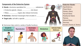
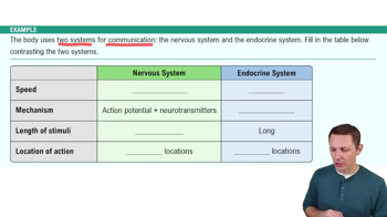
Endocrine system uses hormones released into the bloodstream for long-term regulation. Nervous system uses electrical signals and neurotransmitters for rapid, localized communication.
Controls anterior pituitary via releasing/inhibiting hormones, directly releases ADH and oxytocin through posterior pituitary, and controls sympathetic output to adrenal medulla.
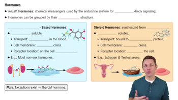
Amino acid derivatives, peptide hormones, and lipid derivatives (steroid hormones).
Amino acid-based hormones bind membrane receptors and use second messengers; steroid hormones enter cells and activate genes directly.
A cell that has the specific receptor to bind and respond to a particular hormone.
Examples: Hypothalamus (ADH, oxytocin), Pituitary (GH, TSH, ACTH), Thyroid (T3, T4, calcitonin), Parathyroids (PTH), Adrenal glands (epinephrine, cortisol), Pancreas (insulin, glucagon).
ACTH → adrenal cortex; TSH → thyroid; GH → liver/somatic tissues; PRL → mammary glands; FSH/LH → gonads; ADH → kidneys; Oxytocin → uterus and mammary glands.
Excess in childhood causes gigantism, excess in adults causes acromegaly, deficiency in childhood causes dwarfism.
Parathyroid hormone (PTH) raises blood Ca2+; calcitonin lowers blood Ca2+.
Growth hormone (via somatomedins), thyroid hormones (T3/T4), insulin, and sex hormones support normal growth and development.
1. Alarm (fight or flight), 2. Resistance (long-term adaptation), 3. Exhaustion (system failure).
Transport gases, nutrients, hormones, wastes; regulate pH and ions; restrict fluid loss; defend body; stabilize temperature.
About 50–60% of blood volume, mostly water with proteins (albumin, globulins, fibrinogen), ions, nutrients, gases, hormones, and wastes.
Blood cell formation in red bone marrow from hemocytoblasts differentiating into myeloid or lymphoid stem cells.
Biconcave discs, no nucleus/mitochondria, lifespan ~120 days, transport O2/CO2 via hemoglobin.
4 globin chains (2 alpha, 2 beta in adults), each with heme containing Fe that binds one O2 molecule.
Stimulated by erythropoietin (EPO) from kidneys in response to low oxygen, promoting RBC production in bone marrow.
Type A: A antigen, anti-B antibody; B: B antigen, anti-A antibody; AB: A and B antigens, no antibodies; O: no antigens, anti-A and anti-B antibodies.
Rh+ has D antigen; Rh− lacks it and can develop anti-Rh antibodies after exposure, causing hemolytic disease in Rh− mothers with Rh+ fetuses.
Neutrophils: bacterial phagocytosis; Lymphocytes: immune response; Monocytes: macrophages; Eosinophils: parasites/allergy; Basophils: inflammation.
1. Vascular spasm, 2. Platelet plug formation, 3. Coagulation forming fibrin clot.
Located in mediastinum; chambers: right atrium/ventricle (pulmonary circuit), left atrium/ventricle (systemic circuit).
Epicardium (outer), myocardium (muscle), endocardium (inner lining).
Venae cavae → right atrium → tricuspid valve → right ventricle → pulmonary valve → lungs → pulmonary veins → left atrium → mitral valve → left ventricle → aortic valve → systemic circulation.
SA node → internodal pathways → AV node → AV bundle → bundle branches → Purkinje fibers.
Atrial systole, ventricular systole (isovolumetric contraction and ejection), ventricular diastole (isovolumetric relaxation and filling).
S1 (lub) = AV valves close; S2 (dub) = semilunar valves close.
\(CO=HR\times SV\), where CO = cardiac output, HR = heart rate, SV = stroke volume.
\(SV=EDV-ESV\). SV increases with preload (EDV), contractility; decreases with afterload.
Sympathetic stimulation increases HR and contractility; parasympathetic stimulation decreases HR.