Skip to main content
Anatomy & Physiology
My Course
Learn
Exam Prep
AI Tutor
Study Guides
Textbook Solutions
Flashcards
Explore
Try the app
My Course
Learn
Exam Prep
AI Tutor
Study Guides
Textbook Solutions
Flashcards
Explore
Try the app
Back
Anatomy & Physiology I: Tissue and Histology Study Guide
You can tap to flip the card.
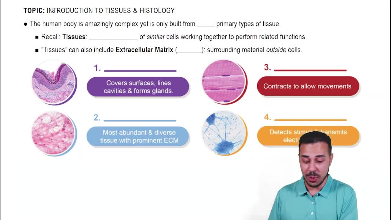
What are the four major tissue types in the human body?
You can tap to flip the card.
👆
What are the four major tissue types in the human body?
Epithelial, connective, muscle, and nervous tissues
.
Track progress
Control buttons has been changed to "navigation" mode.
1/21
Recommended videos
03:32
Map of the Lesson on Tissues
12305
views
548
rank
2
comments
02:52
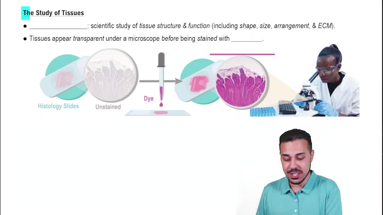
What is Histology?
12417
views
277
rank
04:14
Intro to Human Tissues
20523
views
691
rank
Terms in this set (21)
Hide definitions
What are the four major tissue types in the human body?
Epithelial, connective, muscle, and nervous tissues
.
What is the main characteristic of epithelial tissue surfaces?
Epithelial tissues have a free (apical) surface facing air or fluid-filled spaces called
lumens
and an attached surface on a
basement membrane
.
Where is simple squamous epithelium found and what is its function?
Found in
alveoli of lungs
and
kidney
, it allows for
diffusion and filtration
due to its thin, flat cells.
What distinguishes stratified squamous epithelium, keratinized from non-keratinized?
Keratinized type is found only in
skin
with anucleate surface cells; non-keratinized is found in
esophagus
with surface cells containing nuclei.
What is the function of goblet cells in epithelial tissue?
Goblet cells secrete
mucus
to lubricate and protect epithelial surfaces.
Name the connective tissue type that is most widely distributed and diverse.
Connective tissue
is the most widely distributed and diverse tissue type in the body.
What are the main fiber types found in areolar connective tissue?
Areolar connective tissue contains
collagen fibers
(thick, pink),
elastic fibers
, and
reticular fibers
(both thinner).
How can adipose tissue be identified with different stains?
With H&E stain, fat droplets appear
white
(washed out), while with osmic acid (fat stain), fat droplets stain
black
.
Where is dense regular connective tissue found and what is its fiber arrangement?
Found mainly in
tendons and ligaments
, fibers are tightly packed
collagen arranged in parallel rows
.
What is the key feature of elastic connective tissue?
Contains abundant
elastic fibers
allowing tissues to
stretch and recoil
.
Where is hyaline cartilage located and what are its identifying features?
Located in
trachea, larynx, nose, articular surfaces, ribs, fetal skeleton
; features include
chondrocytes in lacunae
and a
smooth matrix
.
How does fibrocartilage differ from hyaline cartilage?
Fibrocartilage has numerous
collagen fibers
giving a
grainy matrix
appearance, found in
intervertebral discs and symphysis pubis
.
What are the main components of compact bone tissue?
Compact bone consists of
osteons (Haversian systems)
,
lamellae
,
central canals
,
lacunae
with osteocytes, and
canaliculi
.
What is the matrix of blood called and what are its formed elements?
The matrix is
plasma
; formed elements include
erythrocytes
,
leukocytes
(granulocytes and agranulocytes), and
platelets
.
Name the three types of muscle tissue and a key identifying feature of each.
Skeletal:
multinucleate, striated, peripheral nuclei;
Cardiac:
branched, striated, intercalated discs;
Smooth:
spindle-shaped, no striations, single central nucleus.
What unique structure is found only in cardiac muscle tissue?
Intercalated discs
connect cardiac muscle cells and allow synchronized contraction.
What are the two major cell types in nervous tissue?
Neurons
(nerve cells) and
neuroglial cells
(support cells).
What is the function of the myelin sheath in nervous tissue?
The myelin sheath insulates axons to
increase the speed of nerve impulse conduction
.
How can you distinguish an artery from a vein in cross-section?
Arteries have a
thick muscular wall (tunica media)
and smaller lumen; veins have a
thinner wall
and larger lumen.
What is the tunica intima composed of in blood vessels?
The tunica intima consists of
simple squamous epithelium (endothelium)
plus a thin layer of underlying connective tissue.
What is the appearance of a nerve in cross-section?
Nerves have no lumen and show cross-sections of
myelinated axons
, often described as
frothy
in appearance.
 BackBack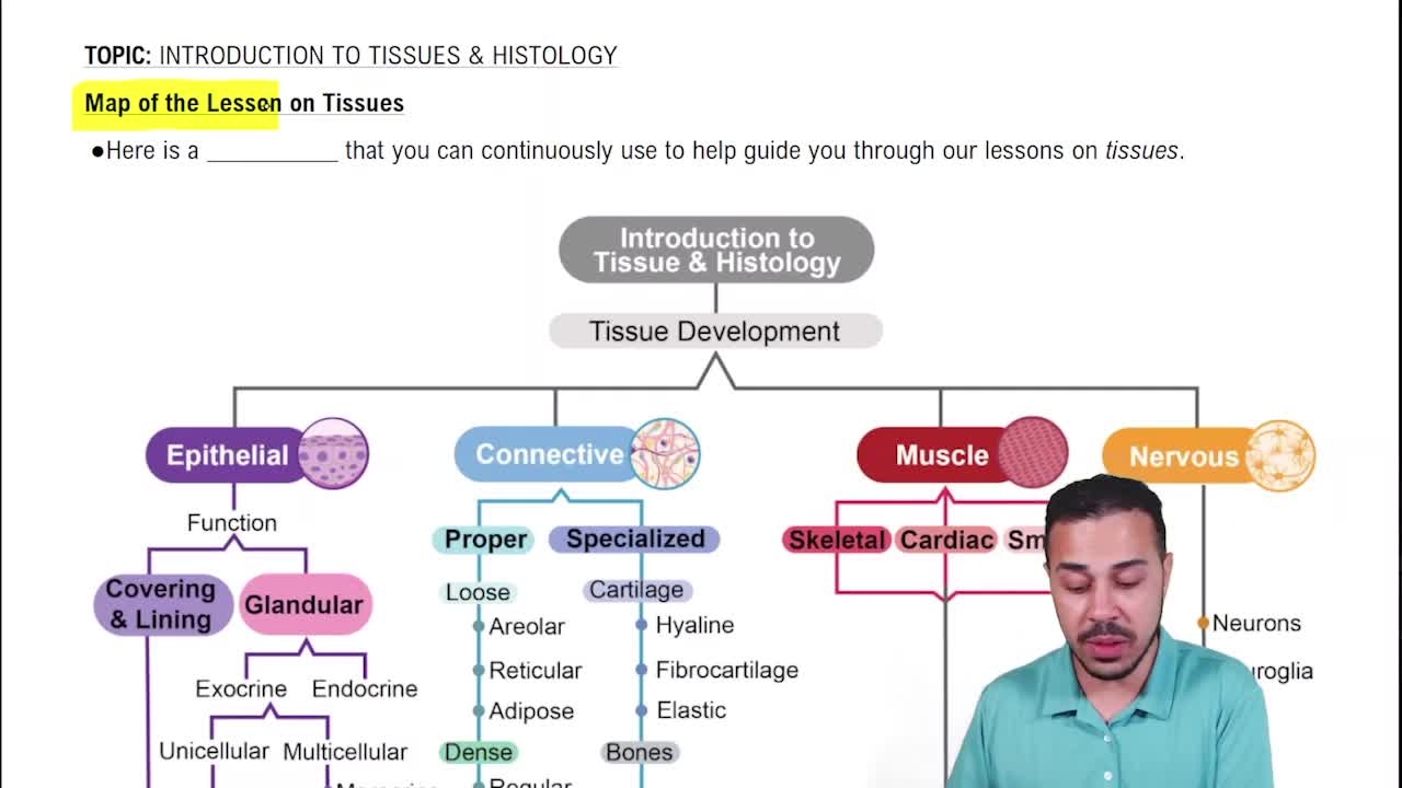
 03:32
03:32