 Back
Anatomy & Physiology: Key Concepts from ANP 1111 Lectures
 03:32

Terms in this set (29)
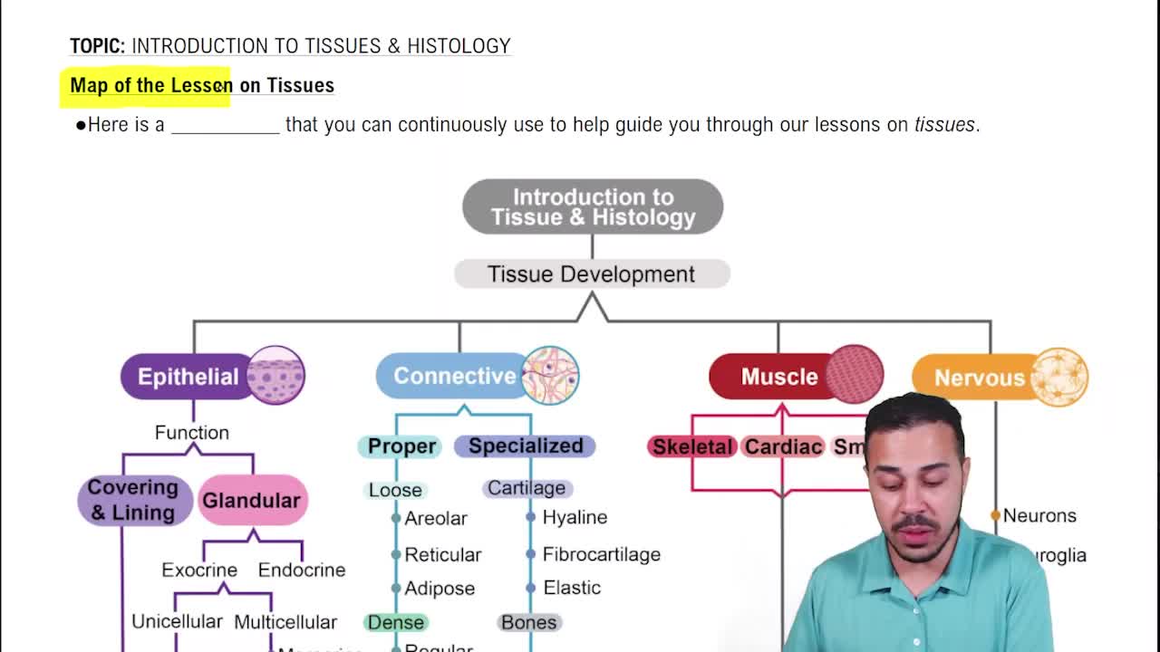
Epithelial tissue forms sheets of cells that cover body surfaces or line cavities, creating boundaries between different environments and performing functions like protection, absorption, filtration, excretion, secretion, and sensory reception.
- Protection
- Absorption
- Filtration
- Excretion
- Secretion
- Sensory reception
Apical surface faces the lumen or external environment and may have microvilli or cilia.
Basal surface attaches to the basement membrane.
- Tight junctions: prevent substances passing between cells.
- Desmosomes: strongly connect adjacent cells.
- Hemidesmosomes: attach cells to the basement membrane.
By cell shape: squamous (flat), cuboidal (cube-shaped), columnar (tall).
By layers: simple (one layer), stratified (multiple layers).
Thin cells allow rapid exchange of gases; found in lungs and parts of the kidney.
Appears stratified due to nuclei at different heights but all cells contact the basement membrane; cilia move mucus; found in respiratory tract (trachea, bronchi).
Specialized stratified epithelium in the urinary bladder that stretches and changes shape as the bladder fills.
Endocrine glands: ductless, secrete hormones into bloodstream.
Exocrine glands: secrete products into ducts leading to body surfaces or cavities.
- Merocrine: secretion via vesicles, cell remains intact.
- Holocrine: entire cell ruptures to release product.
- Apocrine: apical portion of cell pinches off to release product.
- Ground substance/matrix: fills spaces between cells and fibers.
- Fibers: collagen (strength), elastic (stretch), reticular (supportive network).
- Cells: e.g., fibroblasts that produce matrix.
- Areolar: supports epithelia, cushions organs, immune defense.
- Adipose: stores energy, insulates, cushions organs.
- Reticular: forms supportive framework for blood and immune cells.
Dense regular: collagen fibers parallel, resist force in one direction (tendons, ligaments).
Dense irregular: collagen fibers in multiple directions, resist force from many directions (dermis, organ capsules).
Allows tissues to stretch and recoil; found in elastic ligaments and some arteries.
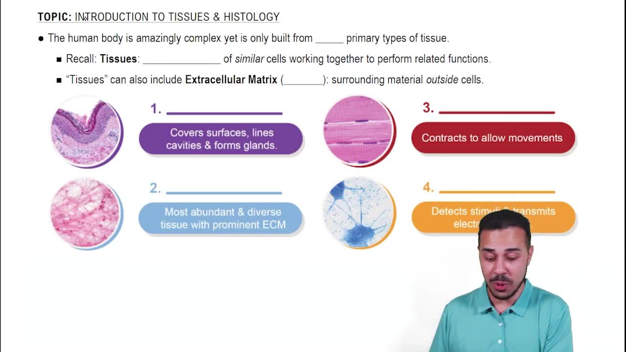
- Nervous: communication.
- Muscle: movement.
- Epithelial: boundaries and coverings.
- Connective: support, protection, binding.
Atoms → Molecules → Organelles → Cells → Tissues → Organs → Organ Systems
Body erect, feet slightly apart, arms at sides, palms facing forward, thumbs pointing away from body; right and left refer to the patient's sides.
- Superior: toward the head.
- Inferior: toward the feet.
- Anterior: toward the front.
- Posterior: toward the back.
- Medial: toward the midline.
- Lateral: away from the midline.
- Sagittal plane: divides body into left and right.
- Frontal (coronal) plane: divides body into front and back.
- Transverse plane: divides body into top and bottom.
Homeostasis maintains stable internal conditions via a control system: Stimulus → Receptor → Control centre → Effector → Response.
Negative feedback: response reverses original change to restore balance.
Positive feedback: response amplifies original change until a specific event stops it.
Cartilage is avascular, flexible yet strong, contains chondroblasts and chondrocytes in lacunae, and has a matrix rich in water and collagen fibers.
- Hyaline: most common, cushioning; found in joints, nose, trachea.
- Elastic: flexible, elastic fibers; external ear, epiglottis.
- Fibrocartilage: strongest, withstands compression; intervertebral discs, knee menisci.
- Support
- Protection
- Movement
- Mineral storage
- Blood cell production
- Fat storage
- Hormone production
- Long: femur, fingers.
- Short: carpals, tarsals.
- Flat: skull, ribs.
- Irregular: vertebrae, hip bones.
An osteon consists of concentric lamellae rings around a central canal containing blood vessels and nerves; osteocytes reside in lacunae connected by canaliculi.
- Osteogenic cells: stem cells producing osteoblasts.
- Osteoblasts: build new bone.
- Osteocytes: maintain bone matrix.
- Osteoclasts: resorb/break down bone.
Thin actin filaments slide over thick myosin filaments, shortening sarcomeres and muscle fibers; filaments themselves do not shorten.
Ca²⁺ binds troponin, moving tropomyosin to expose actin binding sites; ATP binds myosin to detach it from actin and hydrolysis re-energizes myosin heads for repeated cycles.