 Back
Anatomy & Physiology Lecture Test 2 Review
 00:36

Terms in this set (30)
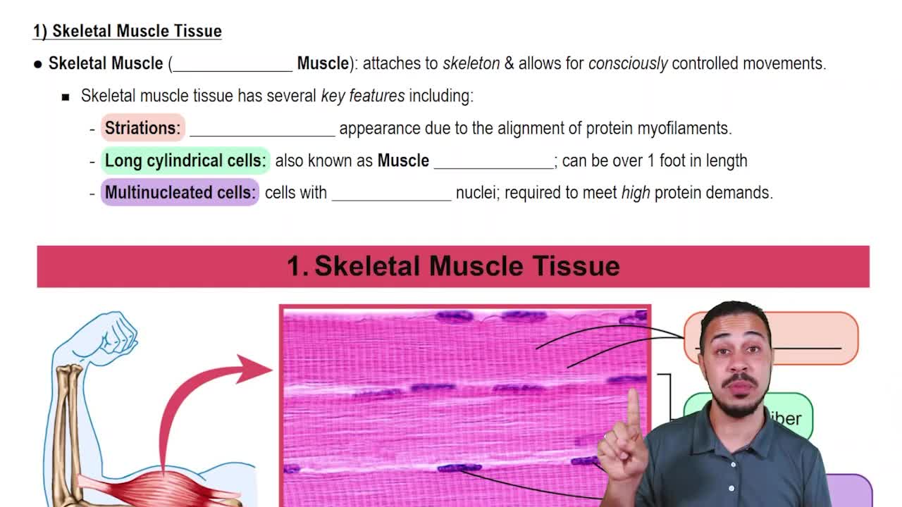
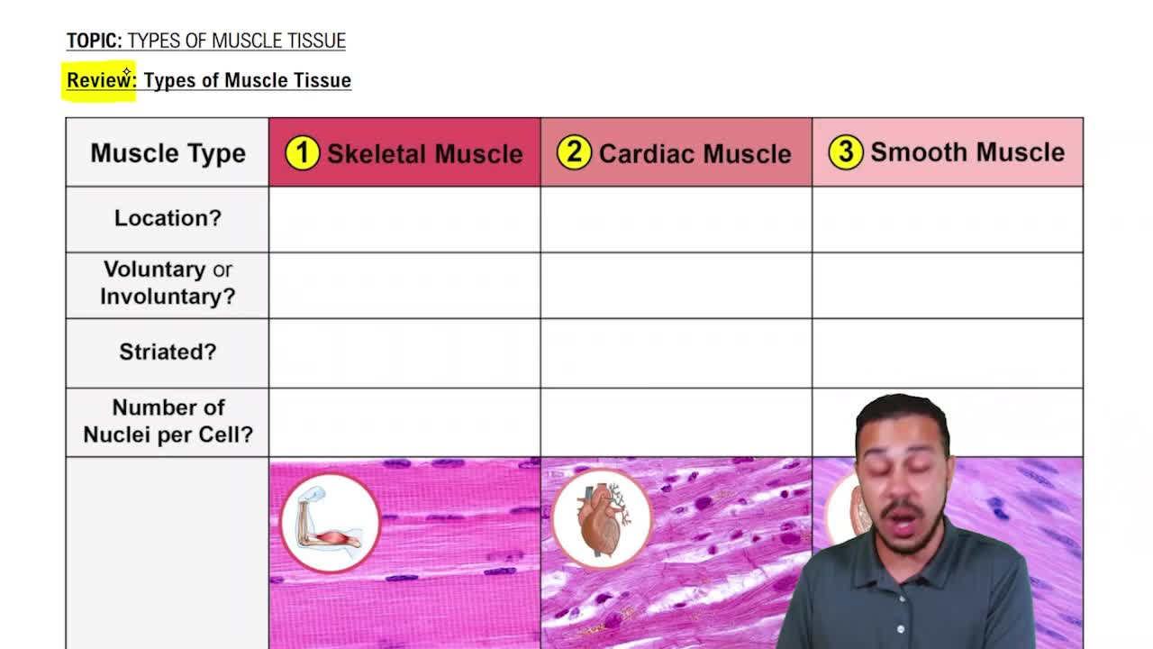
Three types: skeletal (voluntary, striated), cardiac (involuntary, striated), and smooth (involuntary, non-striated).
Synapse where a motor neuron communicates with a skeletal muscle fiber to trigger contraction via neurotransmitter release.
Include flexion, extension, abduction, adduction, rotation, and circumduction, depending on joint type and axis of movement.
Origin is the fixed attachment point; insertion is the movable attachment point of a muscle.
Include diaphysis (shaft), epiphyses (ends), metaphysis (growth plate area), periosteum, compact bone, spongy bone, and medullary cavity.
Osteoblasts build bone, osteocytes maintain bone, and osteoclasts break down bone.
Sensory input, integration, and motor output.
Divided into central nervous system (CNS) and peripheral nervous system (PNS).
Sensory (afferent) division carries signals to CNS; motor (efferent) division carries signals from CNS to effectors.
Somatic (voluntary control of skeletal muscles) and autonomic (involuntary control of smooth, cardiac muscle, glands).
Support, protect, and nourish neurons; maintain homeostasis and form myelin.
Oligodendrocytes form myelin in CNS; Schwann cells form myelin in PNS.
Insulates axons and increases the speed of nerve impulse conduction.
Gaps in the myelin sheath that facilitate rapid impulse conduction via saltatory conduction.
Dendrites receive signals, axon hillock initiates impulses, axon transmits impulses, axon terminals release neurotransmitters.
Irritability: ability to respond to stimuli; conductivity: ability to transmit impulses.
Rapid change in membrane potential caused by ion movement, leading to nerve impulse transmission.
Neurotransmitters like acetylcholine, serotonin, dopamine transmit signals across synaptic cleft to next neuron or effector.
Order: receptor, sensory neuron, integration center, motor neuron, effector.
Somatic reflexes involve skeletal muscles; autonomic reflexes involve smooth or cardiac muscles and glands.
Cerebral hemispheres, diencephalon, brain stem, cerebellum, corpus callosum, and limbic system (hippocampus, amygdala).
Frontal (motor), parietal (sensory), temporal (auditory), occipital (visual), with association areas integrating information.
Thalamus (sensory relay), hypothalamus (homeostasis), and epithalamus (pineal gland).
Midbrain (visual/auditory relay), pons (respiratory control), medulla oblongata (cardiovascular, respiratory centers).
From outer to inner: dura mater, arachnoid mater, pia mater; protect CNS.
Protects brain and spinal cord, circulates nutrients and removes waste; formed by choroid plexus.
Dorsal horns process sensory input; ventral horns control motor output.
12 pairs of cranial nerves with sensory, motor, or mixed functions as per lecture material.
Autonomic controls involuntary effectors; somatic controls voluntary skeletal muscles.
Parasympathetic conserves energy; sympathetic prepares body for 'fight or flight'.