 Back
Anatomy & Physiology: Nervous System and Special Senses
 0:50

Terms in this set (30)
Information gathered by sensory receptors about internal and external changes.
Processing and interpretation of sensory input to decide on a response.
Activation of effector organs (muscles and glands) to produce a response.
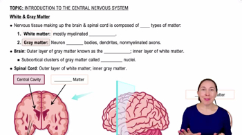
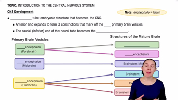
Brain and spinal cord; acts as integration and control center interpreting sensory input and dictating motor output.
Portion of nervous system outside CNS; consists mainly of nerves extending from brain and spinal cord.
Voluntary nervous system controlling conscious skeletal muscle movements.
Involuntary nervous system regulating smooth muscle, cardiac muscle, and glands; has sympathetic and parasympathetic divisions.
Most abundant glial cells; support neurons, control chemical environment, and participate in brain information processing.
Small cells that monitor neurons and can transform to phagocytize microorganisms and debris.
Glial cells that form myelin sheaths around CNS nerve fibers.
Glial cells in PNS that form myelin sheaths and aid regeneration of damaged peripheral nerve fibers.
Whitish, protein-lipid covering that protects and electrically insulates axons, increasing nerve impulse speed.
State where the inside of a neuron is negatively charged relative to the outside.
Decrease in membrane potential making inside less negative, increasing chance of impulse.
Increase in membrane potential making inside more negative, decreasing chance of impulse.
Principal way neurons send signals; involves opening voltage-gated channels and does not decay over distance.
Fast nerve impulse conduction in myelinated axons where impulses jump between nodes of Ranvier.
Junction where neurons communicate with other neurons or effector cells.
Gray matter layer enabling awareness, communication, memory, understanding, and voluntary movement.
Brain structures that regulate muscle movements, filter inappropriate responses, and influence cognition and emotion.
Main visceral control center regulating homeostasis, autonomic nervous system, body temperature, hunger, thirst, and sleep-wake cycles.
Protective coverings of CNS consisting of dura mater, arachnoid mater, and pia mater.
Selective barrier that protects brain by controlling substances passing from blood to neurons.
Twelve pairs of nerves that carry sensory, motor, or both types of information to and from the brain.
Part of ANS that promotes maintenance functions and conserves energy.
Part of ANS that mobilizes the body during activity or stress.
Receptors that bind acetylcholine; include nicotinic (always stimulatory) and muscarinic (excitatory or inhibitory).
Receptors responding to epinephrine and norepinephrine; include alpha (a1, a2) and beta (B1, B2, B3) subtypes with varied effects.
Rods detect dim light and peripheral vision; cones detect bright light and color, concentrated in fovea centralis.
Organ of smell located in the roof of the nasal cavity containing olfactory sensory neurons.