Skip to main content
Anatomy & Physiology
My Course
Learn
Exam Prep
AI Tutor
Study Guides
Textbook Solutions
Flashcards
Explore
Try the app
My Course
Learn
Exam Prep
AI Tutor
Study Guides
Textbook Solutions
Flashcards
Explore
Try the app
Back
Anatomy & Physiology: Nervous System & Special Senses
You can tap to flip the card.
Meninges
You can tap to flip the card.
👆
Meninges
The three protective membranes covering the brain and spinal cord: dura mater, arachnoid mater, and pia mater.
Track progress
Control buttons has been changed to "navigation" mode.
1/31
Recommended videos
0:50
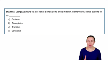
Introduction to the Central Nervous System Example 1
13800
views
256
rank
2
comments
2:41
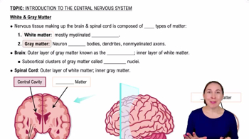
White & Gray Matter
13822
views
249
rank
3:34
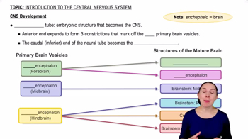
CNS Development
14477
views
290
rank
2
comments
Terms in this set (31)
Hide definitions
Meninges
The three protective membranes covering the brain and spinal cord: dura mater, arachnoid mater, and pia mater.
Epidural space
The space between the dura mater and the vertebral wall, containing fat and blood vessels.
Dura mater
The tough, outermost layer of the meninges that protects the brain and spinal cord.
Arachnoid mater
The middle meningeal layer, a web-like structure that cushions the central nervous system.
Subarachnoid space
The space between the arachnoid mater and pia mater filled with cerebrospinal fluid.
Pia mater
The delicate, innermost meningeal layer that closely adheres to the brain and spinal cord surface.
Central nervous system (CNS)
Comprises the brain and spinal cord, responsible for processing and sending neural signals.
Peripheral nervous system (PNS)
Includes cranial and spinal nerves that connect the CNS to limbs and organs.
Cerebrum
The largest brain part, responsible for voluntary activities, sensory perception, and cognition.
Cerebral cortex
The outer gray matter layer of the cerebrum involved in higher brain functions.
Corpus callosum
A thick band of white matter connecting the two cerebral hemispheres for communication.
Brain lobes
Four main lobes: frontal, parietal, temporal, and occipital, each with specific functions.
Primary motor area
Region of the frontal lobe controlling voluntary muscle movements.
Primary somatosensory area
Region of the parietal lobe processing sensory information from the body.
Hypothalamus
Part of the diencephalon regulating homeostasis, hormone release, and autonomic functions.
Thalamus
Relay center in the diencephalon that processes and transmits sensory information to the cerebral cortex.
Cerebellum
Brain region coordinating voluntary movements, balance, and posture.
Brain stem
Includes midbrain, pons, and medulla oblongata; controls vital functions like breathing and heart rate.
Ventricles of the brain
Four fluid-filled cavities producing and circulating cerebrospinal fluid: lateral, third, and fourth ventricles.
Choroid plexus
Network of cells in ventricles that produces cerebrospinal fluid.
Spinal cord gray matter
Contains neuron cell bodies and is shaped like a butterfly in cross-section.
Spinal cord white matter
Contains myelinated axons forming ascending and descending tracts.
Dorsal root ganglion
Cluster of sensory neuron cell bodies located on the dorsal root of a spinal nerve.
Phrenic nerve
Nerve from the cervical plexus that controls the diaphragm for breathing.
Brachial plexus
Network of nerves supplying the shoulder, arm, and hand.
Sciatic nerve
Largest nerve in the body, originating from the sacral plexus and supplying the lower limb.
Fibrous layer of the eye
Outer layer including the sclera and cornea providing protection and shape.
Vascular layer of the eye
Middle layer containing the choroid, iris, and pupil controlling light entry.
Nervous layer of the eye
Innermost layer, the retina, containing photoreceptors for vision.
Auditory ossicles
Three small bones in the middle ear: malleus, incus, and stapes that transmit sound vibrations.
Vestibulocochlear nerve
Cranial nerve responsible for hearing and balance.
 BackBack
 0:50