 Back
BackAnatomy & Physiology: Nervous System and Vertebral Curvatures
 0:50
0:50
Terms in this set (32)
Primary curvatures are the thoracic and sacral curves present at birth. Secondary curvatures are the cervical and lumbar curves that develop after birth to help with upright posture.
Scoliosis: lateral curvature; Kyphosis: exaggerated thoracic curve; Lordosis: exaggerated lumbar curve.
Includes sensory input, integration, and motor output to control body functions and respond to stimuli.
CNS: brain and spinal cord; PNS: all nerves outside CNS, subdivided into sensory and motor divisions.
Sensory (afferent) carries signals to CNS; Motor (efferent) carries signals from CNS to effectors.
Somatic controls voluntary muscles; Visceral (autonomic) controls involuntary muscles and glands.
Includes receptor level, circuit level, and perceptual level for processing sensory information.
Support neurons: Astrocytes (support, blood-brain barrier), Oligodendrocytes (myelinate CNS), Microglia (immune defense), Ependymal cells (CSF production), Schwann cells (myelinate PNS), Satellite cells (support PNS neurons).
Neurons have a cell body, dendrites (receive signals), and an axon (transmits signals).
Myelin insulates axons to speed signal conduction; formed by Schwann cells in PNS and oligodendrocytes in CNS.
Gaps in myelin sheath where ion channels are concentrated, enabling saltatory conduction of action potentials.
Includes multipolar, bipolar, and unipolar neurons based on number of processes.
Sensory (afferent), motor (efferent), and interneurons based on signal direction and function.
Electrical potential across membrane at rest, typically around \(-70\,mV\), maintained by ion gradients and permeability.
Chemical gradient and electrical gradient for ions, mainly K+ and Na+, maintain resting potential.
Intracellular fluid high in K+, extracellular fluid high in Na+ and Cl-.
Depolarization: membrane potential becomes less negative; repolarization: returns to resting; hyperpolarization: more negative than resting.
Graded potentials vary in magnitude and decay with distance; action potentials are all-or-none, propagate without decay.
Includes resting, depolarization (Na+ channels open), repolarization (K+ channels open), and hyperpolarization phases.
Action potentials propagate along axon by sequential opening of voltage-gated channels, enabling signal transmission.
Absolute refractory: no new AP possible; relative refractory: new AP possible with stronger stimulus.
Junction between neurons where neurotransmitters are released to transmit signals to postsynaptic cells.
Excitatory depolarize postsynaptic membrane; inhibitory hyperpolarize it, affecting likelihood of AP.
Multiple EPSPs and IPSPs combine spatially or temporally to influence postsynaptic neuron firing.
Potentiation increases synaptic strength; presynaptic inhibition reduces neurotransmitter release.
Acetylcholine is a neurotransmitter that binds receptors to excite or inhibit postsynaptic cells, important in muscle activation.
Frontal: motor control, decision making; Parietal: sensory processing; Occipital: vision; Temporal: auditory; Insula: visceral sensation.
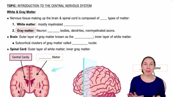
Outer layer of cerebrum responsible for conscious thought, sensory perception, and voluntary motor control.
Association fibers connect areas within same hemisphere; commissural fibers connect hemispheres; projection fibers connect cortex to lower brain/spinal cord.
Regulate voluntary motor control, procedural learning, and movement coordination.
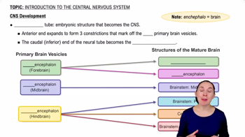
Diencephalon includes thalamus and hypothalamus; thalamus relays sensory and motor signals to cerebral cortex.
Regulates autonomic functions, endocrine system, temperature, hunger, thirst, sleep, and emotional responses.