Skip to main content
Anatomy & Physiology
My Course
Learn
Exam Prep
AI Tutor
Study Guides
Textbook Solutions
Flashcards
Explore
Try the app
My Course
Learn
Exam Prep
AI Tutor
Study Guides
Textbook Solutions
Flashcards
Explore
Try the app
Back
Anatomy & Physiology: Respiratory System
You can tap to flip the card.
What are the main anatomical divisions of the respiratory tract?
You can tap to flip the card.
👆
What are the main anatomical divisions of the respiratory tract?
Upper respiratory tract
includes nasal cavity to larynx;
Lower respiratory tract
includes trachea to alveoli.
Track progress
Control buttons has been changed to "navigation" mode.
1/25
Recommended videos
Guided course
4:28
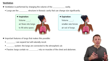
Ventilation
8373
views
195
rank
1
comments
Guided course
5:55
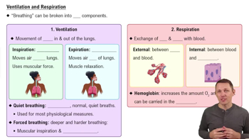
Ventilation and Respiration
12330
views
366
rank
1
comments
Guided course
1:37
Introduction to Lung Physiology Example 1
8381
views
223
rank
1
comments
Terms in this set (25)
Hide definitions
What are the main anatomical divisions of the respiratory tract?
Upper respiratory tract
includes nasal cavity to larynx;
Lower respiratory tract
includes trachea to alveoli.
What are the primary functions of the conducting zone in the respiratory system?
Conducting zone tubes filter, warm, and moisten air; serve as conduits for air from nose to bronchioles.
What structures make up the respiratory zone?
Respiratory zone includes respiratory bronchioles, alveolar ducts, alveolar sacs, and alveoli where gas exchange occurs.
Name the three unpaired cartilages of the larynx.
Thyroid cartilage, cricoid cartilage, and epiglottis.
What is the function of the epiglottis during swallowing?
Epiglottis closes the glottis to prevent food and liquids from entering the larynx and lower respiratory tract.
Describe the histology of the trachea.
Lined with pseudostratified ciliated columnar epithelium and goblet cells; supported by C-shaped hyaline cartilage rings.
What is the carina and its significance?
Last tracheal cartilage ring forming a hook at bifurcation; contains sensory receptors triggering cough reflex.
How do the primary bronchi differ anatomically?
Right primary bronchus is wider, shorter, and more vertical; left is narrower, longer, and more horizontal due to heart position.
What changes occur in the bronchial tree as airways get smaller?
Cartilage changes from rings to plates, epithelium thins from pseudostratified to columnar, and smooth muscle amount increases.
What cell types are found in alveoli and their functions?
Type I alveolar cells for gas diffusion, Type II alveolar cells produce surfactant, alveolar macrophages remove debris.
What is the respiratory membrane?
Thin barrier formed by fused alveolar and capillary walls allowing gas exchange between air and blood.
What are the pleurae and their functions?
Parietal and visceral pleurae are serous membranes enclosing lungs; pleural fluid lubricates and reduces friction during breathing.
Explain Boyle's law in relation to pulmonary ventilation.
At constant temperature, pressure and volume of gas are inversely related; increasing lung volume decreases pressure, causing air inflow.
What are the three pressures influencing pulmonary ventilation?
Atmospheric pressure, intrapulmonary (alveolar) pressure, and intrapleural pressure.
How do inspiratory muscles increase lung volume?
Diaphragm contracts downward; external intercostals elevate ribs, expanding thoracic cavity and lungs.
What is the role of surfactant in alveoli?
Reduces surface tension to prevent alveolar collapse and increases pulmonary compliance.
Define tidal volume (TV) and inspiratory reserve volume (IRV).
TV is air inspired or expired during normal breathing (~500 ml); IRV is additional air forcibly inspired after TV.
What is the oxygen-hemoglobin dissociation curve?
Graph showing relationship between blood PO2 and hemoglobin saturation; demonstrates oxygen loading and unloading.
How does increased temperature affect hemoglobin's oxygen affinity?
Decreases affinity, promoting oxygen unloading to tissues.
Describe the three main ways carbon dioxide is transported in blood.
Dissolved in plasma (~7-10%), bound to hemoglobin as carbaminohemoglobin (~20%), and as bicarbonate ions (~70%).
What is the function of central chemoreceptors in ventilation control?
Detect changes in CO2 and H+ in cerebrospinal fluid and adjust ventilation rate to maintain blood pH.
What is the effect of hyperventilation on blood pH?
Decreases CO2, reduces carbonic acid, raises pH causing respiratory alkalosis.
What are the main features of chronic obstructive pulmonary disease (COPD)?
Persistent airway obstruction, increased airway resistance, and impaired expiration; includes emphysema and chronic bronchitis.
What is the significance of the pleural cavity pressure being lower than atmospheric pressure?
Creates a suction effect that keeps lungs inflated and prevents collapse.
How does ventilation-perfusion coupling optimize gas exchange?
Blood flow is directed to well-ventilated alveoli; airflow is directed to well-perfused areas.
 BackBack
 4:28