 Back
Anatomy & Physiology: Sensory Systems and Special Senses
 5:05

Terms in this set (24)
Sensory transduction is the process by which sensory receptors convert a stimulus (like light, sound, or chemicals) into an electrical signal (action potential) that the nervous system can interpret.
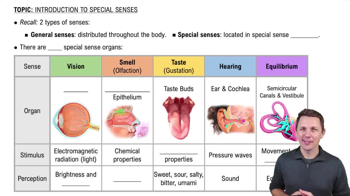
General senses detect touch, pressure, pain, temperature, and proprioception and are located throughout the body. Special senses include smell, taste, vision, hearing, and balance, and have specialized organs.
Olfactory receptors are located in the olfactory epithelium on the roof of the nasal cavity.
Odorants bind to specific receptor proteins on olfactory receptor cells, triggering a signal transduction pathway that generates an action potential.
Action potentials travel from olfactory receptors through the olfactory nerve (CN I) to the olfactory bulb, then to the olfactory cortex and other brain areas.
Taste buds are located mainly on the tongue's papillae and contain gustatory receptor cells that detect chemicals in food.
Signals from taste buds travel via the facial (CN VII), glossopharyngeal (CN IX), and vagus (CN X) nerves to the gustatory cortex.
Sweet (sugars), salty (metal ions), sour (acids), bitter (alkaloids), and umami (amino acids like glutamate).
Cornea (light entry), lens (focuses light), iris (controls pupil size), retina (photoreceptors), sclera (protective outer layer), and optic nerve (transmits visual info).
Fibrous layer: sclera and cornea; vascular layer: choroid, ciliary body, iris; inner layer: retina.
The retina contains photoreceptors (rods and cones), bipolar cells, and ganglion cells that process and transmit visual information.
Light changes the shape of photopigments in rods and cones, triggering a chemical cascade that alters membrane potential and generates signals.
Rods detect low light and peripheral vision; cones detect color and detail in bright light.
Light passes cornea → aqueous humor → lens → vitreous humor → retina; signals travel from retina via optic nerve to the visual cortex.
Light adaptation is the eye adjusting to bright light; dark adaptation is adjusting to low light, involving photopigment regeneration.
Outer ear: pinna, external auditory canal, tympanic membrane; Middle ear: ossicles (malleus, incus, stapes), pharyngotympanic tube.
It connects the middle ear to the throat and helps equalize air pressure on both sides of the tympanic membrane.
Cochlea detects sound; vestibule senses static equilibrium; semicircular canals detect dynamic equilibrium (head rotation).
Sound waves travel through outer ear, vibrate tympanic membrane, ossicles amplify vibrations, cochlea converts vibrations to neural signals.
Sound → external auditory canal → tympanic membrane → ossicles → cochlea → auditory nerve → auditory cortex.
Pitch depends on which part of the cochlea is stimulated; loudness depends on the amplitude of vibrations.
Static equilibrium senses head position relative to gravity; dynamic equilibrium senses head movement and acceleration.
Maculae are sensory patches in the vestibule with hair cells and otoliths that detect head position for static equilibrium.
Crista ampullaris are sensory organs in semicircular canals with hair cells that detect rotational head movements for dynamic equilibrium.