 Back
Anatomy & Physiology: Sensory Systems, Nervous System, and Special Senses
 2:47

Terms in this set (77)
Sensation is the detection of stimuli by sensory receptors. Perception is the brain's interpretation of these sensory signals.
- Mechanoreceptors: respond to mechanical pressure or distortion.
- Thermoreceptors: detect temperature changes.
- Photoreceptors: respond to light.
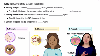
- Chemoreceptors: detect chemical stimuli.
- Nociceptors: sense pain from tissue damage.
- Receptor Level: sensory receptors detect stimuli and transduce signals.
- Circuit Level: pathways transmit impulses to the brain.
- Perceptual Level: brain interprets sensory input.
Transduction is the conversion of a stimulus into an electrical signal by a sensory receptor.
- Stimulus specificity: receptor responds only to certain stimuli.
- Receptive field: area monitored by a receptor.
- Phasic receptors: fast-adapting, respond briefly (e.g., pressure receptors).
- Tonic receptors: slow or non-adapting, respond continuously (e.g., pain receptors).
Somatosensory impulses are delivered to the primary somatosensory cortex in the parietal lobe.
Interpretation occurs at the perceptual level in the cerebral cortex.
Referred pain occurs when pain from internal organs is perceived in somatic areas due to shared neural pathways (e.g., heart attack felt in left arm).
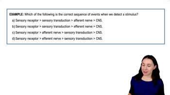
- Sensory (afferent) nerves: carry impulses to the CNS.
- Motor (efferent) nerves: carry impulses from the CNS to effectors.
- Endoneurium: surrounds individual axons.
- Perineurium: surrounds fascicles (bundles of axons).
- Epineurium: surrounds entire nerve.
A mixed nerve contains both sensory and motor fibers; most nerves are mixed to allow two-way communication.
There are 31 pairs: 8 cervical, 12 thoracic, 5 lumbar, 5 sacral, and 1 coccygeal.
Nerves C1–C7 exit above their corresponding vertebrae.
Spinal nerve C8 exits below vertebra C7, unlike C1–C7.
Nerves T1 and below exit below their corresponding vertebrae.
- Dorsal ramus: innervates muscles and skin of the back.
- Ventral ramus: innervates limbs and anterior body wall.
- Meningeal branch: innervates meninges and vertebrae.
A nerve plexus is a network of ventral rami that redistributes fibers to form peripheral nerves, providing redundancy and complex innervation.
- Cervical: phrenic nerve.
- Brachial: median, ulnar, radial nerves.
- Lumbar: femoral nerve.
- Sacral: sciatic nerve.
Called intercostal nerves, they innervate intercostal muscles and skin of the thorax and abdomen.
- Receptor
- Sensory neuron
- Integration center
- Motor neuron
- Effector
Somatic reflexes affect skeletal muscles; autonomic reflexes affect smooth muscle, cardiac muscle, or glands.
The stretch reflex is monosynaptic and ipsilateral, causing muscle contraction; it involves reciprocal inhibition of antagonists.
Stretch reflex is monosynaptic and causes contraction; tendon reflex is polysynaptic and causes muscle relaxation to prevent damage.
Flexor reflex withdraws limb (ipsilateral, polysynaptic); crossed-extensor reflex stabilizes opposite limb (contralateral).
SNS targets skeletal muscles; ANS targets smooth muscle, cardiac muscle, and glands.
SNS: single motor neuron; ANS: two-neuron chain (preganglionic and postganglionic neurons).
Preganglionic neuron cell bodies are in CNS; postganglionic neuron cell bodies are in autonomic ganglia.
- SNS motor neurons release acetylcholine (ACh).
- ANS preganglionic fibers release ACh.
- ANS postganglionic fibers release norepinephrine (sympathetic) or ACh (parasympathetic).
Effect depends on receptor type on target organ, not just neurotransmitter.
- Parasympathetic: "rest and digest"; conserves energy.
- Sympathetic: "fight or flight"; prepares body for stress.
- Parasympathetic: craniosacral origin, long preganglionic, short postganglionic fibers, ganglia near organs.
- Sympathetic: thoracolumbar origin, short preganglionic, long postganglionic fibers, ganglia near spinal cord.
Oculomotor (III), Facial (VII), Glossopharyngeal (IX), Vagus (X).
Provides about 90% of parasympathetic outflow, innervating thoracic and abdominal organs.
- Cardiac plexus: regulates heart rate.
- Pulmonary plexus: controls lungs.
- Esophageal plexus: controls esophagus.
- Abdominal aortic plexus: innervates abdominal organs.
Form pelvic splanchnic nerves that innervate pelvic organs like bladder, reproductive organs, and distal colon.
Paired ganglia located lateral to the vertebral column, forming the sympathetic chain.
- Synapse at same level ganglion.
- Ascend or descend to another ganglion.
- Pass through to collateral ganglia.
Celiac, superior mesenteric, and inferior mesenteric ganglia.
Preganglionic fibers synapse directly on adrenal medulla cells, which secrete epinephrine and norepinephrine into the bloodstream.
Cholinergic fibers release acetylcholine; adrenergic fibers release norepinephrine.
- Nicotinic receptors: found on postganglionic neurons; always stimulatory.
- Muscarinic receptors: found on target organs; can be stimulatory or inhibitory.
- Alpha receptors: usually stimulatory.
- Beta receptors: usually inhibitory but can be stimulatory depending on subtype.
Hypothalamus, brainstem, and spinal cord regulate autonomic functions.
- Lacrimal gland: produces tears.
- Eyelids: protect eye.
- Conjunctiva: lubricates eye surface.
- Extrinsic muscles: move eye.
Tears flow from lacrimal gland → lacrimal ducts → eye surface → lacrimal puncta → lacrimal canaliculi → lacrimal sac → nasolacrimal duct → nasal cavity.
Tears cleanse, protect against infection, and provide nutrients to the cornea.
- Sclera: opaque, protective white outer layer.
- Cornea: transparent, allows light entry and refracts light.
- Choroid: provides blood supply.
- Ciliary body: controls lens shape via ciliary muscle and produces aqueous humor.
- Iris: controls pupil size for light regulation.
- Sphincter pupillae: constricts pupil (parasympathetic).
- Dilator pupillae: dilates pupil (sympathetic).
- Rods: sensitive to low light, no color, high sensitivity, low acuity, located peripherally.
- Cones: color vision, high acuity, less sensitive, concentrated in fovea.
Produced by ciliary body → flows through posterior chamber → pupil → anterior chamber → drains via scleral venous sinus.
Transparent, flexible structure that focuses light onto the retina by changing shape.
Emmetropic vision is focused at 20 feet or more. For close objects, the eye accommodates by lens thickening, pupil constriction, and convergence of the eyes.
Light causes photoreceptors to hyperpolarize, reducing neurotransmitter release and signaling the presence of light.
Light adaptation is rapid adjustment to bright light; dark adaptation is slower due to regeneration of photopigments.
Light → photoreceptors → bipolar cells → ganglion cells → optic nerve → optic chiasm → optic tract → lateral geniculate nucleus → visual cortex in occipital lobe.
Both use chemoreceptors to detect chemical stimuli.
They regenerate olfactory receptor neurons throughout life.
Odorants dissolve in mucus, bind receptors on olfactory neurons, triggering signal transduction.
Olfactory adaptation reduces receptor sensitivity after prolonged exposure.
Olfactory neurons → olfactory bulb → olfactory cortex (temporal lobe) → limbic system and hypothalamus.
Taste buds contain gustatory cells with hairs that detect dissolved chemicals; saliva dissolves tastants for detection.
Sweet, sour, salty, bitter, umami.
Gustatory cells → cranial nerves (VII, IX, X) → medulla → thalamus → gustatory cortex.
Mechanoreceptors in the cochlea detect sound vibrations.
- External ear: auricle, external acoustic meatus.
- Middle ear: tympanic membrane, auditory ossicles.
- Inner ear: cochlea, vestibule, semicircular canals.
Transmit and amplify sound vibrations from tympanic membrane to oval window.
Membranous labyrinth lies within the bony labyrinth and contains endolymph; bony labyrinth contains perilymph.
- Scala vestibuli and scala tympani contain perilymph.
- Scala media contains endolymph and the organ of Corti.
Movement bends hair cell stereocilia, opening ion channels and generating receptor potentials.
Inner hair cells transmit sound signals; outer hair cells amplify and fine-tune sound.
Cochlear nerve → brainstem nuclei → inferior colliculus → medial geniculate nucleus → auditory cortex (temporal lobe).
Vestibule (utricle and saccule) detects linear acceleration; semicircular canals detect rotational movement.
Linear acceleration and head position relative to gravity.
Rotational acceleration of the head.
These areas coordinate balance and posture reflexes rather than conscious perception.