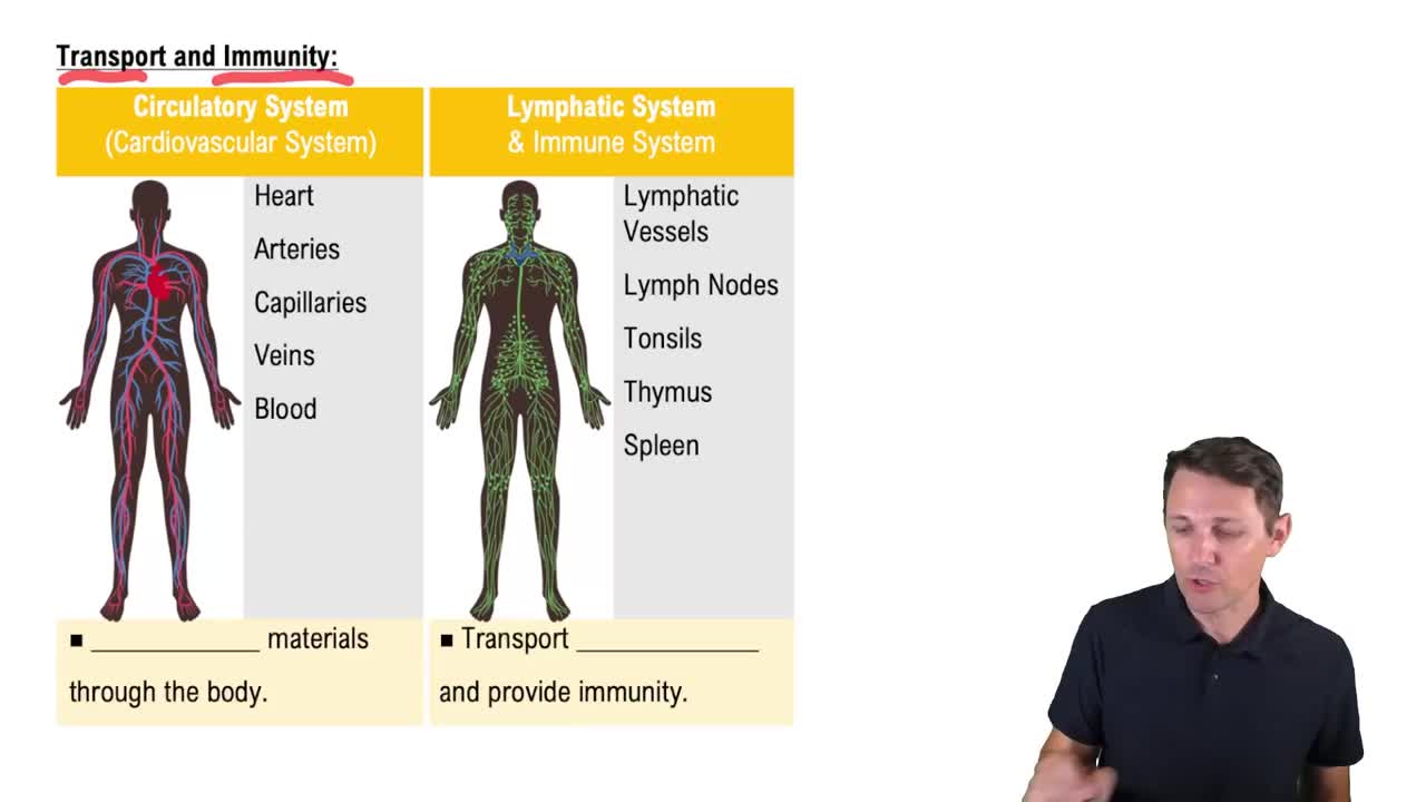
What are the two main circuits of the cardiovascular system?
Pulmonary circuit carries blood to and from the lungs for gas exchange. Systemic circuit carries blood to and from the rest of the body.
What are the three types of blood vessels and their functions?
Arteries carry blood away from the heart. Veins return blood to the heart. Capillaries connect arteries and veins and allow exchange of gases, nutrients, and wastes.
Name the four chambers of the heart and their primary functions.
Right atrium receives blood from systemic circuit. Right ventricle pumps blood into pulmonary circuit. Left atrium receives blood from pulmonary circuit. Left ventricle pumps blood into systemic circuit.
What is the pericardium and its layers?
The pericardium surrounds the heart and consists of an outer fibrous layer and an inner serous layer, which has a parietal layer and a visceral layer (epicardium). The pericardial cavity contains fluid to reduce friction.
What causes pericarditis and what is cardiac tamponade?
Pericarditis is inflammation of the pericardium caused by pathogens. Cardiac tamponade is restricted heart movement due to excess fluid in the pericardial cavity.
What are the sulci of the heart and their significance?
Sulci are grooves on the heart surface containing fat and blood vessels. The coronary sulcus separates atria and ventricles; anterior and posterior interventricular sulci separate left and right ventricles.
Describe the three layers of the heart wall.
Epicardium (visceral serous pericardium) covers the heart surface. Myocardium is cardiac muscle tissue. Endocardium lines inner heart surfaces with simple squamous epithelium.
What is the cardiac skeleton and its functions?
The cardiac skeleton is dense elastic tissue that encircles heart valves and bases of great vessels. It stabilizes valves, supports muscle cells, and electrically insulates atria from ventricles.
What separates the chambers of the heart internally?
The interatrial septum separates the atria, and the interventricular septum separates the ventricles. The interventricular septum is thicker.
What are the atrioventricular (AV) valves and their function?
The AV valves (tricuspid on right, mitral/bicuspid on left) are fibrous flaps between atria and ventricles that allow one-way blood flow from atria to ventricles.
What are the semilunar valves and their role?
The semilunar valves (pulmonary and aortic) prevent backflow of blood from arteries into ventricles during ventricular relaxation.
What vessels bring blood to the right atrium?
The superior vena cava brings blood from head, neck, upper limbs, and chest. The inferior vena cava brings blood from trunk, viscera, and lower limbs.
What is the foramen ovale and its fate after birth?
The foramen ovale is an opening in the fetal interatrial septum allowing blood flow between atria. It closes at birth and becomes the fossa ovalis.
How do the chordae tendineae and papillary muscles function?
Chordae tendineae attach AV valve cusps to papillary muscles, preventing valve prolapse and backflow during ventricular contraction.
What is the moderator band and where is it found?
The moderator band is a muscular ridge in the right ventricle that conducts contraction stimulus to papillary muscles.
What are the major coronary arteries and their supply areas?
The right coronary artery supplies right atrium, parts of ventricles, and conduction system. The left coronary artery supplies left atrium, left ventricle, and interventricular septum.
What is coronary artery disease (CAD)?
CAD is partial or complete blockage of coronary arteries, often due to atherosclerotic plaques, reducing blood flow and cardiac performance.
What is angina pectoris?
Angina pectoris is chest pain caused by temporary ischemia during increased heart workload, often a symptom of CAD.
What causes a myocardial infarction (heart attack)?
Myocardial infarction occurs when coronary circulation is blocked, causing cardiac muscle death and formation of a nonfunctional infarct area.
What is the role of the sinoatrial (SA) node?
The SA node is the heart's primary pacemaker, initiating action potentials at 60–100 beats per minute to establish sinus rhythm.
Describe the conduction pathway of the heart's electrical impulse.
Impulse starts at SA node, spreads through atria to AV node, delays 100 msec, travels via AV bundle, bundle branches, Purkinje fibers, causing ventricular contraction.
What are the components of an electrocardiogram (ECG)?
The ECG includes the P wave (atrial depolarization), QRS complex (ventricular depolarization), and T wave (ventricular repolarization).
What is the refractory period in cardiac contractile cells?
The absolute refractory period (~200 msec) prevents response to stimuli; the relative refractory period (~50 msec) allows response only to strong stimuli, preventing tetany.
How does calcium contribute to cardiac muscle contraction?
Extracellular Ca2+ enters during plateau phase, triggering release of more Ca2+ from sarcoplasmic reticulum, enabling contraction by interacting with contractile proteins.
What are the phases of the cardiac cycle?
The cardiac cycle includes atrial systole, atrial diastole, ventricular systole, and ventricular diastole, involving contraction and relaxation phases.
What causes the heart sounds S1 and S2?
S1 is caused by closure of AV valves; S2 is caused by closure of semilunar valves during the cardiac cycle.
How is cardiac output (CO) calculated?
CO = Heart Rate (HR) × Stroke Volume (SV), where SV = End-Diastolic Volume (EDV) – End-Systolic Volume (ESV).
What factors affect heart rate?
Heart rate is influenced by autonomic nervous system activity, circulating hormones, and reflexes responding to blood pressure and chemical changes.
What is the Frank–Starling principle?
As EDV increases, the stroke volume increases due to greater ventricular stretching and stronger contraction.
 Back
 02:29