 Back
Anatomy & Physiology: Tissue Structure and Function
 03:32

Terms in this set (21)
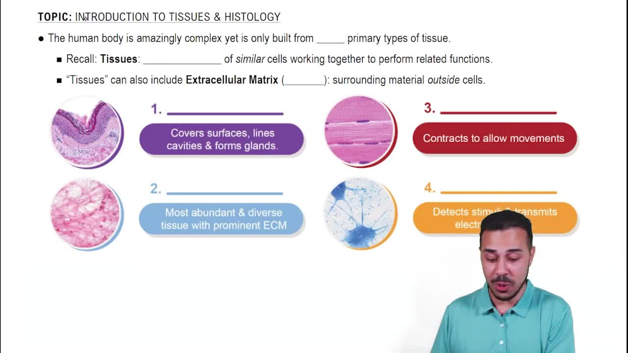
Cells, tissue fluid, and fibers.
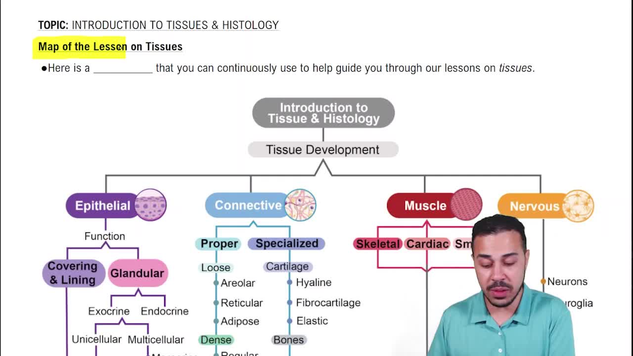
Epithelial, connective, nervous, and muscle tissues.
Cytology studies individual cells, while histology examines tissues composed of multiple cells.
The science of the cause of disease and infection.
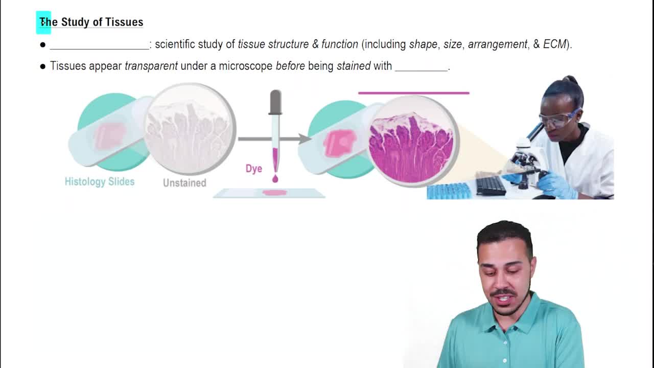
The microscopic examination of tissues to study disease-related changes in cells and tissue structure.
Fixing (preserving), sectioning (thin slicing), and staining (enhancing contrast).
To enhance contrast so structures are more visible under the microscope.
Light microscopy uses colored dyes for staining.
Electron microscopy uses heavy metal coatings to enhance image contrast.
Protection by forming sheets of cells covering surfaces like skin and nails.
Support by surrounding cells with a nonliving extracellular matrix.
Cells with the ability to contract, e.g., skeletal, cardiac, and visceral muscles.
To detect sensations, process information, and control movement and body functions.
Cell membrane, , polarity (apical and basal), and lateral surface features.
Adhesion proteins, membrane contours, and special cell junctions.
They close off intercellular space to prevent molecules from passing between cells.
At the apical region of most epithelial types.
They act as anchoring junctions linking transmembrane proteins and help tissues that stretch.
They form passageways allowing small molecules and electrical signals to move between adjacent cells.
Proteins secreted by epithelial tissues including type IV collagen, laminin, nidogen/entactin, and heparan sulfate proteoglycans.
Provides structural stability, selective permeability, cell signaling, and tissue organization.