Skip to main content
Anatomy & Physiology
My Course
Learn
Exam Prep
AI Tutor
Study Guides
Textbook Solutions
Flashcards
Explore
Try the app
My Course
Learn
Exam Prep
AI Tutor
Study Guides
Textbook Solutions
Flashcards
Explore
Try the app
Back
Anatomy & Physiology: Tissues, Integumentary and Digestive System lab 3 & lab 4
You can tap to flip the card.
What are the four main types of animal tissues?
You can tap to flip the card.
👆
What are the four main types of animal tissues?
Epithelial, connective, muscle, and nervous tissues
.
Track progress
Control buttons has been changed to "navigation" mode.
1/25
Recommended videos
03:32
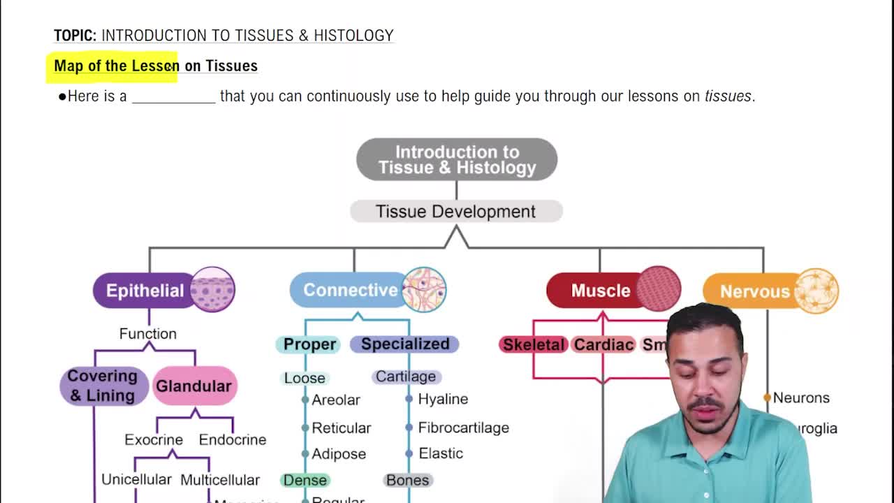
Map of the Lesson on Tissues
14917
views
680
rank
2
comments
02:52
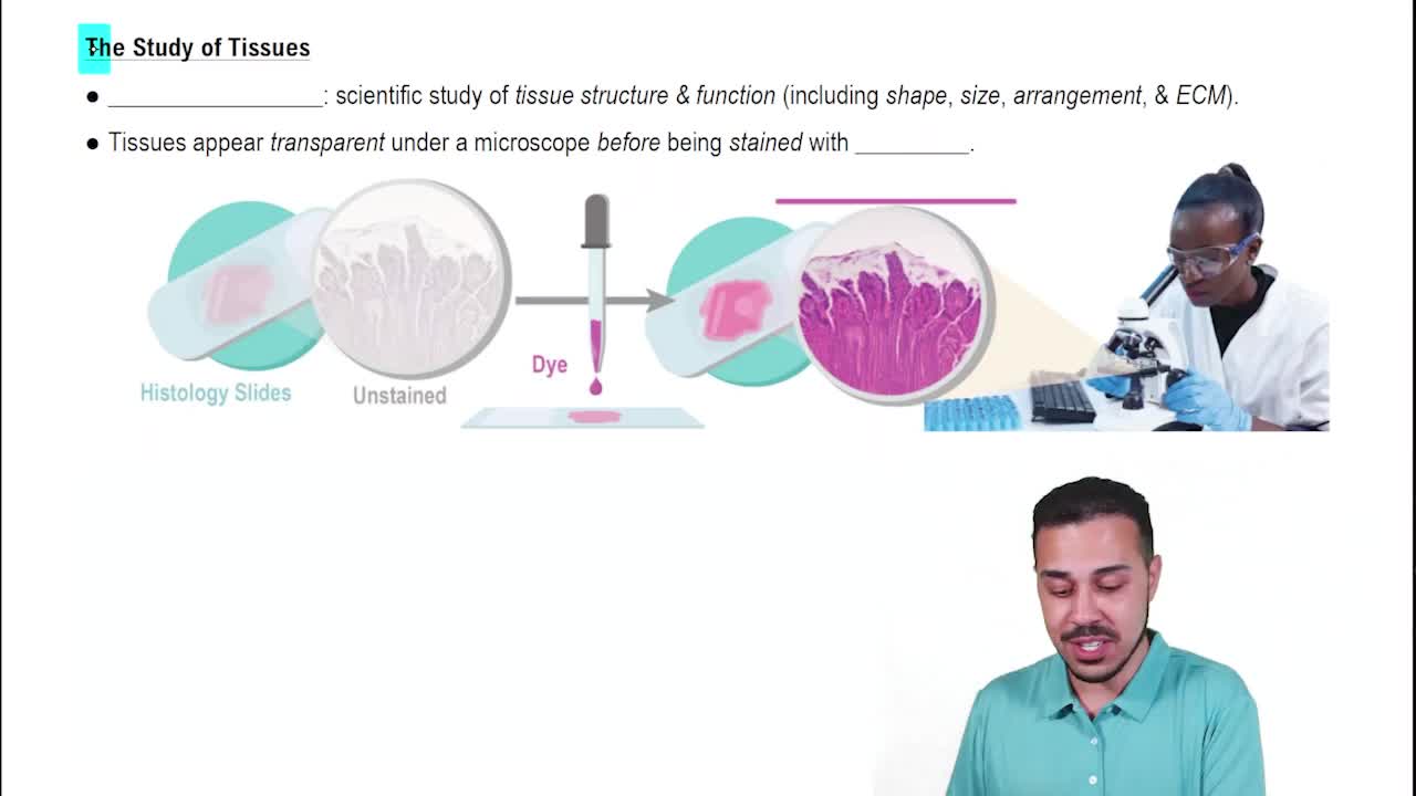
What is Histology?
14995
views
339
rank
04:14
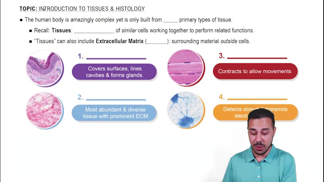
Intro to Human Tissues
24378
views
882
rank
Terms in this set (25)
Hide definitions
What are the four main types of animal tissues?
Epithelial, connective, muscle, and nervous tissues
.
How is epithelial tissue classified?
By
cell shape
(squamous, cuboidal, columnar, transitional) and
number of layers
(simple, stratified, pseudostratified).
What is the main non-living component of connective tissue?
The
matrix
, consisting of fibers and ground substance secreted by cells.
Name the three types of muscle tissue.
Skeletal, cardiac, and smooth muscle
, differing in location, structure, and control.
What are the two layers of the skin (integumentary system)?
The
epidermis
(epithelial tissue) and the
dermis
(connective tissue).
What protein makes keratinocytes tough and water-resistant?
Keratin
.
List the layers of the epidermis from superficial to deep.
Stratum
corneum
,
lucidum
(only in thick skin),
granulosum
,
spinosum
, and
basale
.
What type of connective tissue is the dermis mainly composed of?
Dense, irregular connective tissue rich in
collagen
.
Name two types of sudoriferous (sweat) glands and their locations.
Eccrine glands
found in thick and thin skin;
apocrine glands
associated with hair follicles in axillary and pubertal hair regions.
What is the hypodermis composed of and is it part of the skin?
Composed mainly of
adipose tissue
; it is
not
technically part of the skin.
What is the function of simple squamous epithelial tissue in the body?
Allows
diffusion and filtration
; found lining blood vessels and alveoli.
What connective tissue type provides support and stores energy as fat?
Adipose tissue
.
What are chondrocytes and where are they found?
Cells found in
cartilage
that reside in spaces called
lacunae
.
Describe the structure and function of skeletal muscle tissue.
Long, cylindrical, striated fibers with multiple peripheral nuclei; responsible for
voluntary movement
.
What distinguishes cardiac muscle tissue histologically?
Short, branched, striated fibers with a single central nucleus and
intercalated discs
.
Where is smooth muscle tissue found and what is its function?
Walls of hollow organs; controls
involuntary movements
like food propulsion and blood flow regulation.
What are the main components of nervous tissue?
Neurons and neuroglial cells.
What are the major functions of the integumentary system?
Protection, thermoregulation, and sensation
.
What is the role of melanocytes in the epidermis?
Produce
melanin
, the pigment responsible for skin color and UV protection.
What is the function of the arrector pili muscle?
Causes hair to stand up, producing 'goosebumps' for thermoregulation.
What are the main layers of the digestive tract wall?
Mucosa, submucosa, muscularis, and serosa (or adventitia)
.
What is the function of villi and lacteals in the small intestine?
Villi increase surface area for nutrient absorption; lacteals transport dietary lipids via lymph.
What is the unique muscular feature of the stomach compared to other GI organs?
The stomach has an additional
oblique muscle layer
besides longitudinal and circular layers.
What is the peritoneum and its two major parts?
A serous membrane; the
parietal peritoneum
lines the abdominal cavity, and the
visceral peritoneum
(serosa) covers organs.
Name the major folds of the peritoneum and their functions.
Greater omentum (fat storage), lesser omentum (supports stomach), falciform ligament (supports liver), mesocolon (supports colon), mesentery (supports small intestine).
 BackBack
 03:32