Skip to main content
Anatomy & Physiology
My Course
Learn
Exam Prep
AI Tutor
Study Guides
Textbook Solutions
Flashcards
Explore
Try the app
My Course
Learn
Exam Prep
AI Tutor
Study Guides
Textbook Solutions
Flashcards
Explore
Try the app
Back
AP Chapter 4 part 1
You can tap to flip the card.
Basal lamina
You can tap to flip the card.
👆
Basal lamina
A supporting sheet consisting of proteins secreted by epithelial cells, part of the basement membrane.
Track progress
Control buttons has been changed to "navigation" mode.
1/25
Recommended videos
07:25
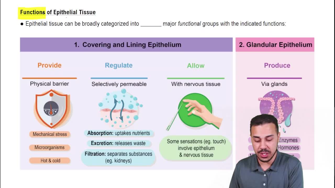
Functions of Epithelial Tissue
16808
views
358
rank
03:40
Intro to Epithelia
19901
views
477
rank
05:24
Introduction to Epithelial Tissue Example 1
13059
views
265
rank
1
comments
Terms in this set (25)
Hide definitions
Basal lamina
A supporting sheet consisting of proteins secreted by epithelial cells, part of the basement membrane.
Five distinguishing characteristics of epithelial tissue
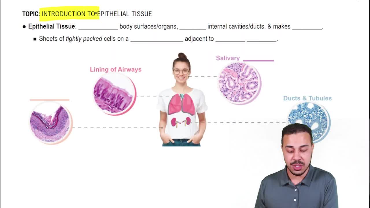
1. Cells bound tightly in sheets 2. Apical and basal surfaces 3. Cells rest on basement membrane 4. Avascular but innervated 5. Capable of rapid cell division.
Apical surface of epithelial cells
The surface facing the exposed outer or inner surface of the organ.
Basal surface of epithelial cells
The surface attached to underlying connective tissue by the basement membrane.
Basement membrane composition
Made up of basal lamina and reticular lamina; reinforces epithelial sheet and resists stretching and tearing.
Simple squamous epithelium function and location
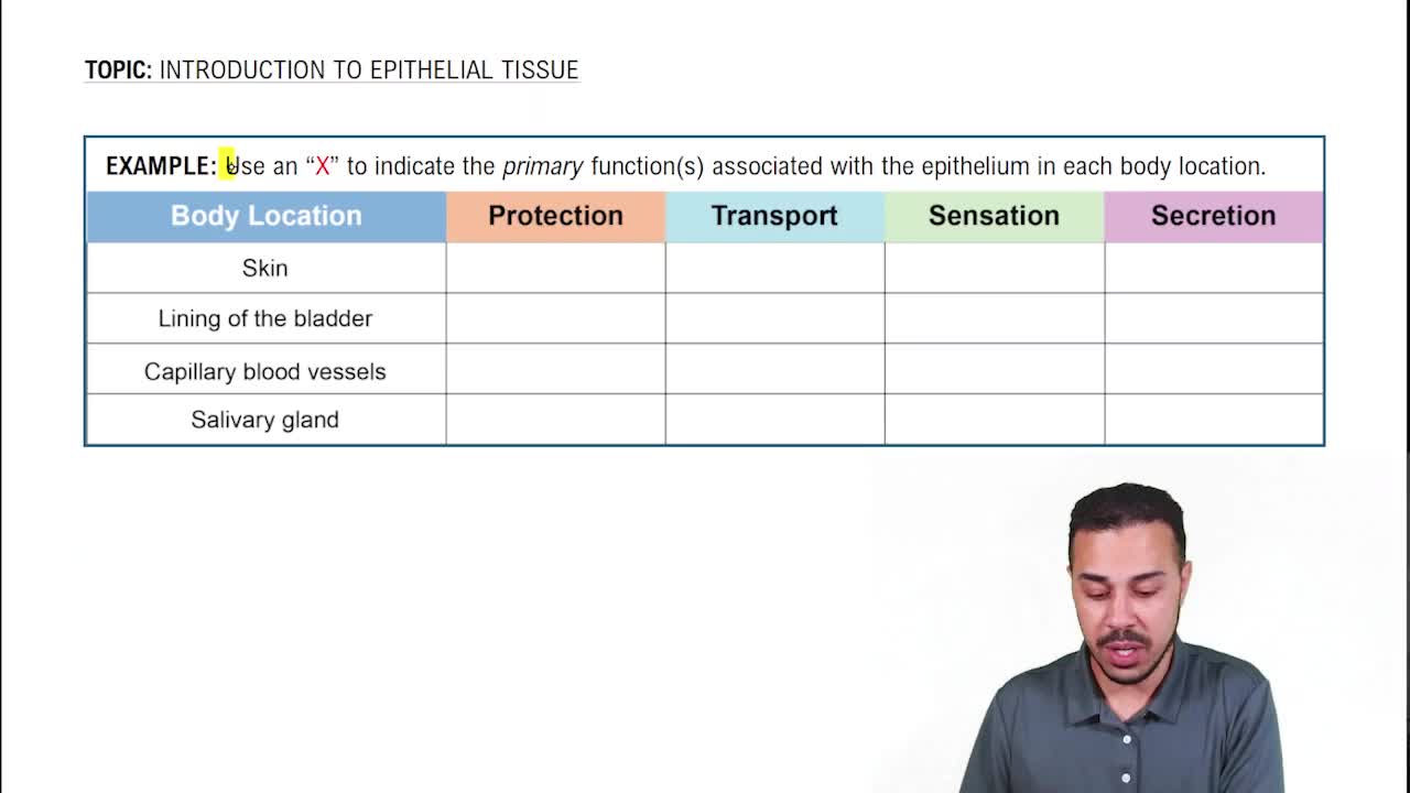
Forms thin membranes for passage of materials; found in air sacs of lungs and lining of heart and blood vessels.
Simple cuboidal epithelium function and location
Adapted for secretion and absorption; found in kidney tubules and pancreatic ducts.
Simple columnar epithelium function and location
Absorbs nutrients and secretes mucus; lines most digestive organs and some exocrine glands.
Pseudostratified columnar epithelium features
Nuclei at varying heights, cells originate from basement membrane, some reach apical surface; secretes and propels mucus.
Pseudostratified columnar epithelium location
Lines trachea, bronchi, and parts of nasal cavity.
Stratified squamous epithelium function and location
Protects against abrasion, drying, and infection; keratinized type in skin epidermis, non-keratinized in mucous membranes.
Transitional epithelium function and location
Allows stretching; found lining urinary bladder, urethra, and ureters.
Microvilli
Finger-like projections forming the brush border; increase surface area for absorption.
Cilia
Short, numerous hair-like appendages that move substances across the cell surface; found in respiratory tract and oviduct.
Keratin
A protein that protects epithelial cells; found in skin, mouth, esophagus, vagina, rectum, hair, nails, and horns.
Difference between endocrine and exocrine glands
Endocrine glands are ductless and secrete hormones into blood; exocrine glands have ducts and secrete onto surfaces or cavities.
Examples of endocrine glands
Pineal gland, pancreas, ovaries, testes, thyroid gland, adrenal gland.
Examples of exocrine glands
Mucous, sweat, oil, salivary glands, liver, pancreas, mammary glands.
Multicellular exocrine gland structure
Composed of a duct and secretory unit, often surrounded by connective tissue forming a capsule and lobes.
Classification of exocrine glands by duct structure
Simple glands have unbranched ducts; compound glands have branched ducts.
Classification of exocrine glands by secretory unit shape
Tubular glands have tube-shaped secretory cells; alveolar glands have sac-shaped secretory cells; tubuloalveolar glands have both.
Merocrine mode of secretion
Secretory vesicles release products by exocytosis without cell damage (e.g., sweat glands, pancreas).
Apocrine mode of secretion
Apical portion of cell pinches off to release secretion; seen in mammary glands.
Holocrine mode of secretion
Entire cell ruptures to release secretion; seen in sebaceous (oil) glands.
Carcinoma
Cancer originating in epithelial tissue; accounts for 80-90% of adult cancers including lung, breast, prostate, and bowel.
 BackBack
 07:25