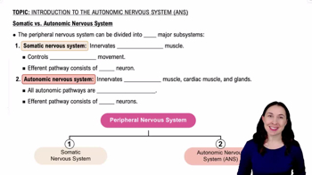
The Nervous System has 2 main divisions: Central Nervous System (CNS) and Peripheral Nervous System (PNS). The PNS further divides into the Somatic Nervous System (SNS) and Autonomic Nervous System (ANS).
Divisions of the Autonomic Nervous System (ANS)
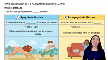
The ANS has two divisions: Sympathetic (fight or flight) and Parasympathetic (rest and digest).
Somatic Nervous System Control
Motor neurons of the CNS exert direct control over skeletal muscles via Lower Motor Neurons (LMN) and Upper Motor Neurons (UMN).
Visceral Reflex Arc Components
Made of 2 motor neurons: Preganglionic neurons (in brainstem/spinal cord) and Ganglionic neurons (in autonomic ganglia) that innervate cardiac muscle, glands, and fat cells.
Enteric Nervous System
A nerve network in the walls of the digestive tract controlled by the ANS and visceral reflexes, containing about 1 million neurons.
Sympathetic Division Location and Fiber Lengths
Also called thoracolumbar division (T1-L2). Has short preganglionic fibers and long postganglionic fibers.
Functions of the Sympathetic Division
Increases tissue metabolism, mental alertness, respiratory function, heart rate, blood pressure, and activates sweat glands; decreases digestive and urinary functions.
Sympathetic Chain Ganglia
Also called paravertebral ganglia; paired chains on both sides of the vertebral column controlling body walls, thoracic cavity, head, and limbs.
Collateral Ganglia
Also called prevertebral ganglia; located anterior to the vertebral column, innervating tissues and organs in the abdominopelvic cavity.
Adrenal Medullae Role
Specialized ganglionic neurons in adrenal glands that release neurotransmitters (norepinephrine, epinephrine) into the bloodstream affecting target cells body-wide.
White Ramus vs Gray Ramus
White ramus: myelinated preganglionic fibers (T1-L2 only). Gray ramus: unmyelinated postganglionic fibers, present in all spinal nerves.
Horner’s Syndrome
Damage to thoracic/cervical sympathetic pathways causing loss of sympathetic control of the face, resulting in pupil constriction and flushed face due to parasympathetic dominance.
Parasympathetic Division Location and Fiber Lengths
Also called craniosacral division; has long preganglionic fibers and short postganglionic fibers. Preganglionic fibers originate in brainstem and sacral spinal cord.
Functions of the Parasympathetic Division
Decreases metabolic activity, heart rate, and blood pressure; increases digestive secretions, blood flow to digestive tract, urination, defecation, and sexual arousal.
Parasympathetic Cranial Nerves
Preganglionic fibers are part of cranial nerves III (oculomotor), VII (facial), IX (glossopharyngeal), and X (vagus), with the vagus providing 75% of parasympathetic innervation.
Neurotransmitters of the ANS
Two main types: Cholinergic (use acetylcholine) and Adrenergic (use norepinephrine or epinephrine).
Cholinergic Receptors
Two types: Nicotinic receptors (excitatory, found on ganglion cells and neuromuscular junctions) and Muscarinic receptors (excitatory or inhibitory, found on target organs).
Adrenergic Receptors
Two main types: Alpha (α) receptors (α1 excitatory, α2 inhibitory) and Beta (β) receptors (β1 excitatory, β2 inhibitory, β3 stimulates lipolysis).
Dual Innervation and Autonomic Tone
Most visceral organs receive input from both sympathetic and parasympathetic divisions; balance between them is called autonomic tone.
Visceral Reflex Example
Pupil size is controlled by sympathetic (dilation) and parasympathetic (constriction) reflexes, modulated by the hypothalamus and higher brain centers.
Types of Mechanoreceptors
Include tactile receptors (touch, pressure), baroreceptors (pressure changes in vessels), and proprioceptors (joint and muscle position).
Types of Tactile Receptors
Six types: Free nerve endings, Root hair plexus, Merkel discs, Meissner's corpuscles, Pacinian corpuscles, and Ruffini corpuscles, each sensitive to different touch or pressure stimuli.
Alzheimer’s Disease Characteristics
Progressive loss of higher cerebellar functions causing senile dementia; associated with plaques, neurofibrillary tangles, and reduced neurotransmitter production.
Effects of Aging on the Nervous System
Includes brain size and weight reduction, neuron loss, decreased blood flow, fewer synapses, and slower motor skills.
 Back
Back
 1:10
1:10