Skip to main content
Anatomy & Physiology
My Course
Learn
Exam Prep
AI Tutor
Study Guides
Textbook Solutions
Flashcards
Explore
Try the app
My Course
Learn
Exam Prep
AI Tutor
Study Guides
Textbook Solutions
Flashcards
Explore
Try the app
Back
Brain and Cranial Nerves - Anatomy & Physiology
You can tap to flip the card.
Major divisions of the brain
You can tap to flip the card.
👆
Major divisions of the brain
Medulla oblongata
,
Pons
,
Mesencephalon (midbrain)
,
Diencephalon
,
Cerebellum
, and
Cerebrum
.
Track progress
Control buttons has been changed to "navigation" mode.
1/31
Recommended videos
Guided course
0:50
Introduction to the Central Nervous System Example 1
14095
views
263
rank
2
comments
Guided course
2:41
White & Gray Matter
14082
views
256
rank
Guided course
3:34
CNS Development
14688
views
295
rank
2
comments
Terms in this set (31)
Hide definitions
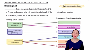
Major divisions of the brain
Medulla oblongata
,
Pons
,
Mesencephalon (midbrain)
,
Diencephalon
,
Cerebellum
, and
Cerebrum
.
Functions of the cerebrum
Responsible for
conscious thought processes
,
intellectual functions
,
memory storage and processing
, and
regulation of skeletal muscle contractions
.
Role of the thalamus
Acts as a
relay and processing center for sensory information
before it reaches the cerebrum.
Functions of the hypothalamus
Controls
emotions
,
autonomic functions
, and
hormone production
; links nervous and endocrine systems.
Functions of the mesencephalon (midbrain)
Processes
visual and auditory data
, generates
reflexive somatic motor responses
, and maintains
consciousness
.
Functions of the cerebellum
Coordinates
complex somatic motor patterns
and adjusts output of other motor centers to maintain
balance and posture
.
Functions of the pons
Relays sensory information to the cerebellum and thalamus; contains
somatic and visceral motor centers
.
Functions of the medulla oblongata
Relays sensory information to the thalamus and brainstem; contains
autonomic centers
regulating cardiovascular, respiratory, and digestive activities.
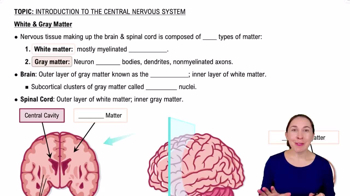
Gray vs. white matter in the brainstem
Gray matter
is inner and surrounds ventricles;
white matter
is outer and consists of axon tracts.
Three layers of cranial meninges
Dura mater
(outer),
arachnoid mater
(middle), and
pia mater
(inner).
Dura mater structure
Two fibrous layers:
endosteal/periosteal
(outer, fused to skull) and
meningeal
(inner), with dural sinuses between.
Meningeal folds and their locations
Falx cerebri
between hemispheres,
tentorium cerebelli
separates cerebrum and cerebellum,
falx cerebelli
divides cerebellar hemispheres,
diaphragma sellae
anchors dura to sphenoid.
Arachnoid mater features
Delicate membrane between dura and pia mater; contains
arachnoid granulations
that allow CSF to enter venous circulation.
Pia mater characteristics
Tightly adheres to brain surface and sulci; highly vascular and anchored by astrocytes.
Ventricles of the brain
Four CSF-filled cavities: two lateral ventricles, third ventricle in diencephalon, and fourth ventricle between pons and cerebellum.
Cerebrospinal fluid (CSF) functions
Fills subarachnoid space, cushions brain, transports nutrients and waste, and is produced by the choroid plexus.
Choroid plexus role
Specialized ependymal cells and fenestrated capillaries produce CSF by active and passive transport.
Blood-brain barrier (BBB) components
Formed by
tight junctions
between endothelial cells, supported by astrocyte end-feet, controlling substance passage to neurons.
Medulla oblongata reflex centers
Cardiovascular centers
regulate heart rate and blood flow;
respiratory rhythmicity centers
set breathing rate.
Pons respiratory centers
Pneumotaxic
and
apneustic centers
modify respiratory output from the medulla oblongata.
Cranial nerves overview
Twelve pairs numbered I to XII; sensory, motor, or both; originate from brain regions like medulla, pons, or mesencephalon.
Olfactory nerve (I) function and pathway
Sensory nerve for smell; receptors in nasal epithelium send signals through cribriform plate to olfactory bulb.
Optic nerve (II) function and pathway
Sensory nerve for vision; fibers cross at optic chiasm, then project to lateral geniculate nucleus and visual cortex.
Oculomotor (III) and Trochlear (IV) nerves
Motor nerves controlling most eye muscles; originate in mesencephalon and pass through superior orbital fissure.
Trigeminal nerve (V) branches and functions
Three branches: ophthalmic, maxillary, mandibular; sensory from face and motor for mastication muscles.
Facial nerve (VII) functions
Sensory for taste and proprioception; motor for facial expression, tear, and salivary glands.
Vestibulocochlear nerve (VIII) functions
Special sensory for balance (vestibular branch) and hearing (cochlear branch).
Glossopharyngeal nerve (IX) functions
Sensory for taste; motor for swallowing muscles and salivary glands; passes through jugular foramen.
Vagus nerve (X) functions
Extensive sensory and motor functions primarily for visceral organs, including respiratory, cardiovascular, and digestive systems.
Accessory nerve (XI) functions
Motor nerve controlling muscles of palate, pharynx, larynx, sternocleidomastoid, and trapezius.
Hypoglossal nerve (XII) functions
Motor nerve controlling voluntary tongue movements; originates in medulla oblongata.
 BackBack
 0:50