 Back
Central Nervous System - Anatomy & Physiology
 0:50

Terms in this set (27)
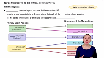
The brain and spinal cord are the major components of the CNS, responsible for receiving information, integrating it, and directing responses.
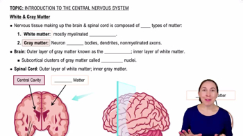
Gray matter contains neuron cell bodies, dendrites, and unmyelinated processes for integration and processing. White matter consists mostly of myelinated axons for communication between regions.
Gray matter forms a central H-shaped region, surrounded by white matter on the outside.
Gray matter forms the outer cortex and deep nuclei, while white matter lies internally.
Cortex: outer layer of gray matter; nucleus: deep cluster of neuron cell bodies; tract: bundle of axons within the CNS.
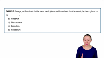
Midbrain → pons → medulla oblongata.
Regulates cardiac activity, vasomotor tone, respiratory rhythm, and reflexes like swallowing, coughing, sneezing, and hiccupping.
Relay information from the cerebral cortex to the cerebellum via the middle cerebellar peduncles.
Superior colliculi mediate visual reflexes; inferior colliculi mediate auditory reflexes.
Contains pigmented neurons linked to basal nuclei and voluntary movement; loss of dopamine here is associated with Parkinson disease.
1. Receives motor plan from cerebral cortex.
2. Compares plan with current body position.
3. Sends corrective feedback to adjust motor commands.
Thalamus, hypothalamus, and epithalamus surround the third ventricle.
Acts as a relay gateway for sensory and other signals traveling to the cerebral cortex.
Autonomic control, emotional responses, thermoregulation, hunger/thirst, behavior, sleep–wake cycle, endocrine regulation, and memory involvement.
Melatonin is secreted and helps regulate the circadian rhythm.
Fissure: deep groove; sulcus: shallow groove; gyrus: raised ridge between sulci.
Hidden deep within the lateral sulcus of the cerebrum.
Commissural fibers connect hemispheres; association fibers connect areas within one hemisphere; projection fibers connect cortex with lower CNS regions.
Extends from the foramen magnum to approximately the L1–L2 vertebral level.
Conus medullaris: tapered end of spinal cord.
Filum terminale: connective tissue anchor to coccyx.
Cauda equina: bundle of spinal nerve roots below conus.
Ascending pathways carry sensory information to the brain.
Descending pathways carry motor commands from brain to spinal cord.
Dura mater → arachnoid mater → subarachnoid space (with CSF) → pia mater.
Produced by choroid plexuses in all four brain ventricles via filtration from blood.
Provides buoyancy and cushioning, nourishes CNS tissue, removes waste, and facilitates chemical signaling.
Lateral ventricles → interventricular foramina → third ventricle → cerebral aqueduct → fourth ventricle → median/lateral apertures → subarachnoid space → arachnoid granulations → dural venous sinuses.
Via arachnoid villi/granulations that drain CSF into dural venous sinuses.
Tight junctions between brain capillary endothelial cells form the BBB. It is selective because it allows oxygen and nutrients through but blocks many toxins and pathogens.