Back

ch 9

Control buttons has been changed to "navigation" mode.
1/18
  • Differences between skeletal, cardiac, and smooth muscle tissue

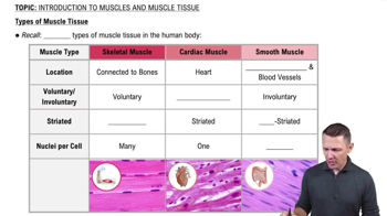
    Skeletal: Striated, voluntary, multinucleated, moves skeleton.
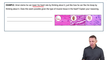
    Cardiac: Striated, involuntary, branching cells with intercalated discs, pumps blood.
    Smooth: Nonstriated, involuntary, spindle-shaped, in hollow organ walls.
  • Major functions of skeletal muscle

    Produce body movement

    Maintain posture

    Support soft tissues

    Guard entrances/exits

    Maintain body temperature

    Store nutrients.

  • Structural hierarchy of skeletal muscle from largest to smallest

    Whole muscle → Muscle fascicle → Muscle fiber (cell) → Myofibril → Sarcomere → Myofilaments (actin and myosin).
  • Three connective tissue layers wrapping a skeletal muscle

    Epimysium: Surrounds entire muscle.

    Perimysium: Surrounds each fascicle.

    Endomysium: Surrounds each muscle fiber.

  • Special terms for muscle cell structures

    Sarcolemma: Plasma membrane.

    Sarcoplasm: Cytoplasm.

    Sarcoplasmic Reticulum (SR): Stores calcium.

    T-tubules: Carry action potentials deep into cell.

  • Boundaries and zones of a sarcomere

    Z lines: Boundaries anchoring thin filaments.

    M line: Center anchoring thick filaments.

    A band: Length of thick filaments.

    I band: Thin filaments only.

    H zone: Thick filaments only.

  • Sarcomere regions that shorten during contraction

    Shorten: I band, H zone, distance between Z lines.

    Stay same: A band length.

  • Sequence of events at the neuromuscular junction (NMJ)

    Action potential arrives → Ca2+ enters axon terminal → ACh released → ACh binds motor end plate → Na+ influx generates muscle action potential → ACh broken down by acetylcholinesterase.
  • What is Excitation–Contraction (E-C) Coupling?

    Action potential travels down T-tubules → triggers Ca2+ release from SR → Ca2+ binds troponin → tropomyosin shifts exposing actin binding sites.
  • Sequence of the Cross-Bridge Cycle

    Ca2+ binds troponin → myosin binds actin → power stroke releases P and ADP → ATP binds myosin causing detachment → ATP hydrolyzed recocks myosin head.
  • Effect of ATP unavailability after death

    Myosin heads cannot detach from actin and Ca2+ cannot be pumped back into SR, causing muscles to lock in contraction (rigor mortis).
  • Definition of a motor unit

    A single motor neuron and all the muscle fibers it innervates; small units for fine control, large units for powerful movements.
  • Difference between wave summation, incomplete tetanus, and complete tetanus

    Wave summation: Twitches add before relaxation.

    Incomplete tetanus: Near-max tension with small relaxations. Complete tetanus: No relaxation, smooth sustained tension.

  • Three types of muscle contractions

    Concentric (Isotonic): Muscle shortens.

    Eccentric (Isotonic): Muscle lengthens while active.

    Isometric: Muscle tension without length change.

  • Three energy systems for ATP regeneration in skeletal muscle

    Phosphocreatine system: Immediate, ~15 sec.

    Anaerobic glycolysis: Fast, no oxygen, 1–2 min.

    Aerobic metabolism: Slow, efficient, uses oxygen, sustained energy.

  • Characteristics of Slow Oxidative (Type I) muscle fibers

    High endurance, small diameter, dark color (myoglobin), many mitochondria, aerobic metabolism.
  • Characteristics of Fast Glycolytic (Type IIb) muscle fibers

    Powerful, large diameter, pale color, low myoglobin, few mitochondria, high glycogen, fatigue quickly.
  • Difference between hypertrophy and atrophy

    Hypertrophy: Increase in muscle fiber size from repeated loading.

    Atrophy: Decrease in muscle size and power from disuse or lack of stimulation.