 Back
Digestive System Anatomy & Physiology
 4:06

Terms in this set (29)
Fuel the body, provide energy, support cell growth, break down food, absorb nutrients, and excrete waste.
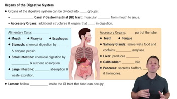
Oral cavity, esophagus, stomach, small intestine, large intestine.
Teeth, tongue, pancreas, gallbladder, liver.
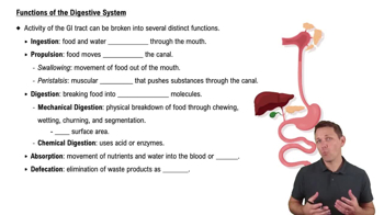
Ingestion, propulsion, mechanical digestion, chemical digestion, absorption, defecation.
Contains most digestive organs; lined by a serous membrane producing peritoneal fluid for lubrication.
Visceral peritoneum (covers organs) and parietal peritoneum (lines cavity walls).
Stabilize and connect organs; double-layered peritoneum suspending intestines and storing fat.
Lesser omentum and falciform ligament.
Contains adipose tissue for insulation and protection of abdominal organs.
Secrete hormones like GIP, GLP-1, and cholecystokinin to regulate digestion.
Epithelial layer and lamina propria (areolar tissue with blood and lymph vessels).
Smooth muscle layers that produce peristalsis to move food along the digestive tract.
Serosa is a protective covering of muscularis externa in most digestive organs; adventitia is connective tissue outer layer in esophagus and rectum.
Stratified squamous epithelium in oral cavity, pharynx, esophagus, anus; simple columnar epithelium in stomach and intestines.
Circular muscles contract behind bolus; longitudinal muscles contract in front to push food forward.
Mixes food with digestive secretions to increase nutrient absorption.
Sensory analysis, lubrication, chewing, and digestion of carbohydrates and proteins.
Parotid (salivary amylase, 25%), sublingual (buffers and lubricant, 5%), submandibular (amylase and mucin, 70%).
99.4% water, 0.6% buffers, electrolytes, enzymes, mucus, and antibodies.
Buccal phase (tongue pushes bolus to pharynx), pharyngeal phase (bolus moves to esophagus), esophageal phase (peristalsis pushes bolus to stomach).
Mechanical digestion, chemical digestion, production of chyme, and intrinsic factor for vitamin B12 absorption.
Parietal cells secrete HCl and intrinsic factor; chief cells produce pepsinogen; mucin cells protect stomach lining.
Pepsinogen is converted to active pepsin by hydrochloric acid (HCl) in the stomach.
Active transport of H+ ions into stomach lumen combines with Cl- to form hydrochloric acid.
Essential glycoprotein for vitamin B12 absorption; deficiency leads to anemia.
G cells produce gastrin to stimulate parietal and chief cells; D cells produce somatostatin to inhibit gastrin.
Cephalic (before food enters stomach), gastric (food in stomach), intestinal (chyme in small intestine).
Low acidity (high pH) increases gastrin release, stimulating more HCl secretion.
High acidity (low pH) inhibits gastrin release, reducing HCl secretion.