 Back
BackDigestive System Anatomy & Physiology (Chapter 23 Part 1)
 4:06
4:06

Terms in this set (21)
Continuous tube through which food passes; includes oral cavity, pharynx, esophagus, stomach, small intestine, and large intestine.
Organs that assist digestion but are not part of the alimentary canal; include teeth, tongue, salivary glands, liver, gallbladder, and pancreas.
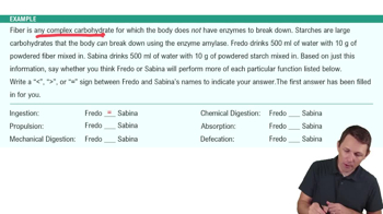
Ingestion, propulsion, mechanical breakdown, digestion, absorption, and defecation.
Largest serous membrane in the body with two layers: parietal peritoneum lining the body wall and visceral peritoneum covering digestive organs.
Folds of visceral peritoneum that hold digestive organs in place, house blood vessels, nerves, lymphatics, and store fat.
Mucosa, submucosa, muscularis externa, and serosa (or adventitia).
Secretion, absorption, and protection; lined by stratified squamous epithelium in mouth/esophagus/anus and simple columnar epithelium elsewhere.
Areolar connective tissue with blood and lymphatic vessels, submucosal glands, and the submucosal plexus of the enteric nervous system.
Two layers of smooth muscle (inner circular, outer longitudinal) responsible for segmentation and peristalsis; stomach has three layers.
Serosa covers intraperitoneal organs (areolar CT), adventitia covers organs outside abdominal cavity (dense CT).
Blood supply and drainage of abdominal digestive organs via branches of abdominal aorta and veins draining into hepatic portal vein.
Veins drain digestive organs and deliver blood to liver for processing before returning to inferior vena cava.
Intrinsic nervous system of GI tract with over 100 million neurons; includes submucosal and myenteric plexuses controlling motility and secretions.
Sympathetic inhibits digestive processes; parasympathetic stimulates digestion.
Site of ingestion, secretion, mechanical breakdown, and propulsion; contains teeth, tongue, and salivary glands.
Hard palate (anterior 2/3) made of stratified squamous epithelium and connective tissue; soft palate (posterior 1/3) made of skeletal muscle with uvula.
Extrinsic muscles control position; intrinsic muscles control shape and size; both assist in chewing and bolus formation.
Parotid (serous cells near ear), submandibular (mostly serous, medial mandible), and sublingual (mostly mucous, under tongue).
97–99.5% water, electrolytes (including bicarbonate), salivary amylase, lysozyme, and secretory IgA.
Primarily parasympathetic via salivatory nucleus and ACh; sympathetic increases mainly mucous secretions.
Located in alveoli of mandible and maxilla, held by periodontal ligament; function in mechanical digestion (mastication) to increase surface area.