 Back
Fundamentals of the Nervous System and Nervous Tissue
 04:36

Terms in this set (23)
Sensing changes (sensory), interpreting changes (integrative), and reacting to changes (motor).
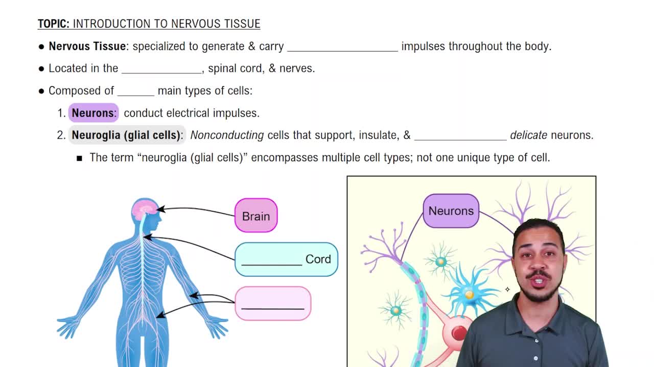
The brain and spinal cord.
Cranial and spinal nerves with sensory (afferent) and motor (efferent) components.
Conducts impulses from skin and special senses to CNS and from CNS to skeletal muscle (voluntary control).
Conveys impulses from CNS to smooth muscle, cardiac muscle, and glands (involuntary control).
Anchor neurons to capillaries, help form the blood-brain barrier, and recapture neurotransmitters.
Wrap cytoplasmic extensions to form myelin sheaths around CNS nerve fibers.
Act as macrophages engulfing microorganisms and dead neural tissue in the CNS.
Form myelin sheaths around PNS nerve fibers and act as phagocytes.
Consists of a cell body (soma), dendrites (input), and usually a single axon (output).
Multipolar: many dendrites, one axon; bipolar: one dendrite, one axon; unipolar: one fused process.
Afferent (sensory): impulses to CNS; association (interneurons): impulses between neurons; efferent (motor): impulses to effectors.
White matter: myelinated axons; gray matter: nerve cell bodies, dendrites, unmyelinated axons, and neuroglia.
Leakage (nongated) channels always open; gated channels open/close in response to stimuli (voltage, chemical, mechanical, light).
Typically -70 mV, caused by ion distribution: more positive sodium ions outside, more negative ions and proteins inside.
Depolarization: membrane potential reverses (+30 mV); repolarization: returns to resting potential (-70 mV) via sodium-potassium pumps.
If threshold is reached, an action potential fires at constant maximum strength; stronger stimuli do not increase impulse size.
Action potentials jump between myelin-covered nodes, speeding impulse conduction in myelinated fibers.
A depolarizing postsynaptic potential that makes the neuron more likely to reach threshold and fire an action potential.
A hyperpolarizing postsynaptic potential that makes the neuron less likely to fire an action potential.
Removed from synaptic cleft by diffusion, enzymatic degradation, or uptake into neurons and glia.
Possible if cell body intact and Schwann cells active; involves Wallerian degeneration and Schwann cell-guided regrowth.
Usually permanent damage; CNS neurons generally do not regenerate after injury.