 Back
BackKidney Anatomy & Nephron Structure

 0:54
0:54
Terms in this set (20)
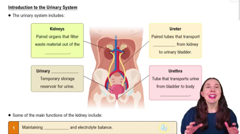
Kidney and ureter are the main external structures visible.
Urinary bladder, trigone, and urethra.
Minor calyx, renal pelvis, major calyx, renal artery, renal vein, and ureter.
Proximal convoluted tubule (PCT), distal convoluted tubule (DCT), descending limb, ascending limb, and nephron loop.
Glomerulus, parietal epithelial cells, and Bowman's capsule.
Afferent arteriole, efferent arteriole, glomerulus, Bowman's capsule, proximal convoluted tubule (PCT), distal convoluted tubule (DCT), loop of Henle, and collecting tubule.
The ureter transports urine from the kidney to the urinary bladder.
The trigone is a triangular region of the bladder base between the openings of the ureters and urethra.
The renal artery carries oxygenated blood to the kidney; the renal vein carries oxygen-depleted blood away.
Minor calyces collect urine from renal pyramids; major calyces collect urine from minor calyces and drain into the renal pelvis.
The PCT reabsorbs water, ions, and nutrients from the filtrate back into the blood.
The DCT further adjusts filtrate composition by selective secretion and reabsorption.
The loop of Henle concentrates urine by reabsorbing water and salts in its descending and ascending limbs.
The collecting tubule collects urine from multiple nephrons and transports it to the renal pelvis.
Parietal epithelial cells form the outer layer of Bowman's capsule.
The glomerulus is a network of capillaries that filters blood plasma into Bowman's capsule.
Afferent arteriole brings blood to the glomerulus; efferent arteriole carries filtered blood away.
Filtrate enters the proximal convoluted tubule (PCT) after Bowman's capsule.
The renal pelvis collects urine from major calyces and funnels it into the ureter.
The nephron filters blood, reabsorbs needed substances, secretes wastes, and forms urine.