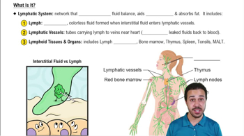
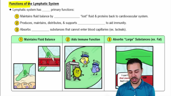
Network of lymphatic vessels, lymph fluid, and lymph nodes that return leaked fluids to blood and cleanse lymph.
Functions of lymphatic vessels
Return interstitial fluid and leaked plasma proteins to blood; lymph flows one-way toward the heart.
What are lacteals?
Specialized lymphatic capillaries in intestinal mucosa that absorb digested fats and deliver fatty lymph (chyle) to the blood.
How is lymph transported through lymphatic vessels?
By skeletal muscle contractions, thoracic pressure changes during breathing, valves preventing backflow, arterial pulsations, and smooth muscle contractions in vessel walls.
Main types of lymphocytes and their roles
T cells moderate immune response and destroy infected cells; B cells produce plasma cells that secrete antibodies.
Role of macrophages in lymphatic system
Phagocytize foreign substances and help activate T cells.
Structure of lymph nodes
Fibrous capsule with trabeculae, cortex with follicles (B cells), deep cortex with T cells, and medullary cords containing B, T, and plasma cells.
How does lymph flow through a lymph node?
Enters via afferent vessels, flows through sinuses to medulla, exits via fewer efferent vessels, allowing immune cells time to act.
Functions of the spleen
Site of lymphocyte proliferation, blood cleansing of aged cells, stores breakdown products of RBCs, platelets, and monocytes.
Unique features of the thymus
Site of T cell maturation, lacks B cells and follicles, most active in childhood, contains thymic corpuscles for regulatory T cell development.
What is MALT and its function?
Mucosa-associated lymphoid tissue protects mucous membranes from pathogens; includes tonsils, Peyer's patches, and appendix.
Two lines of innate defense
1) Surface barriers like skin and mucosae; 2) Internal defenses including phagocytes, natural killer cells, antimicrobial proteins, inflammation, and fever.
How do phagocytes destroy pathogens?
Phagocytes adhere to pathogens, engulf them into phagosomes, fuse with lysosomes to form phagolysosomes, and digest the pathogens.
Role of natural killer (NK) cells
Attack cells lacking 'self' markers, induce apoptosis in cancer and virus-infected cells, and secrete chemicals enhancing inflammation.
Cardinal signs of acute inflammation
Redness, heat, swelling (edema), pain, and impairment of function.
Functions of inflammatory mediators
Dilate arterioles, increase capillary permeability, attract leukocytes, and promote clotting to isolate injured area.
Steps of phagocyte mobilization during inflammation
Leukocytosis, margination, diapedesis, and chemotaxis to the injury site.
How do interferons protect against viruses?
Infected cells secrete interferons that warn neighbors to produce antiviral proteins blocking viral replication and activate NK cells.
What is the complement system?
A group of ~20 blood proteins that enhance inflammation, promote phagocytosis, and cause cell lysis of pathogens.
Three pathways of complement activation
Classical (antibody-mediated), lectin (lectin binding to sugars), and alternative (spontaneous activation).
Role of membrane attack complex (MAC)
Formed by complement proteins inserting into pathogen membranes, creating pores that cause cell lysis.
 Back
 3:32