 Back
BackMicroscopy, Epithelial and Connective Tissue Types in Anatomy & Physiology

 03:32
03:32
Terms in this set (25)
Ocular lens, nosepiece, objective lenses, mechanical stage, condenser, iris, illumination, arm, fine adjustment, coarse adjustment, base, and stage control.
Total magnification = power of ocular lens (usually 10) × power of objective lens.
Resolution is the ability to distinguish between two close objects, with a minimum of 0.2 micrometers for light microscopes.
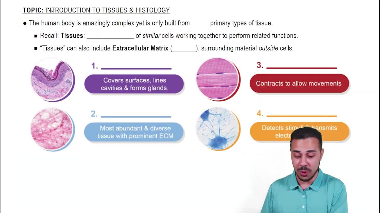
Epithelial, connective, muscle, and nerve tissues.
Simple (one layer) and stratified (multiple layers).
Squamous (flat), cuboidal (cube-shaped), and columnar (tall and column-shaped).
Single layer of flattened cells; allows diffusion and filtration; secretes lubricating substances.
Kidney glomeruli, air sacs of lungs, lining of heart, blood vessels, and ventral body cavity.
Single layer of cube-like cells with large spherical nuclei; functions in secretion and absorption.
Kidney tubules, ducts and secretory portions of small glands, ovary surface.
Single layer of tall cells with round to oval nuclei; absorption and secretion of mucus and enzymes; may have cilia.
Digestive tract lining, gallbladder, excretory ducts of glands, uterine tubes, and uterus.
Single layer of cells of differing heights; secretes mucus and propels it with cilia.
Nonciliated type in male sperm-carrying ducts; ciliated type lines trachea and upper respiratory tract.
Thick membrane with several cell layers; protects underlying tissues in areas subject to abrasion.
Nonkeratinized type forms moist linings of esophagus, mouth, vagina; keratinized type forms epidermis.
Resembles both stratified squamous and cuboidal; stretches readily to allow organ expansion.
Lines ureters, bladder, and part of urethra.
Gel-like matrix with fibroblasts, macrophages, mast cells; wraps and cushions organs; holds tissue fluid.
Closely packed adipocytes with nucleus pushed aside; provides reserve food fuel, insulation, and protection.
Network of reticular fibers forming soft internal skeleton; supports white blood cells, mast cells, and macrophages.
Primarily parallel collagen fibers; attaches muscles to bones or muscles; withstands tensile stress in one direction.
Irregularly arranged collagen fibers; withstands tension from many directions; provides structural strength.
Amorphous but firm matrix with collagen fibers; supports and reinforces; cushions and resists compressive stress.
Embryonic skeleton, ends of long bones, costal cartilages, nose, trachea, and larynx.