Back

Muscle Structure and Function - Anatomy & Physiology

Control buttons has been changed to "navigation" mode.
1/20
  • Three types of muscle tissue

    Skeletal muscle: striated, voluntary, attached to bones.
    Cardiac muscle: striated, involuntary, found in heart.
    Smooth muscle: non-striated, involuntary, found in internal organs.
  • Skeletal muscle fiber characteristics

    Long, cylindrical, multinucleate cells with striations. Contain sarcolemma, sarcoplasm, sarcoplasmic reticulum, myofibrils, mitochondria, and glycogen granules.
  • Muscle fiber components

    Muscle fibers contain myofibrils composed of thin (actin) and thick (myosin) filaments, organized into sarcomeres.
  • Sarcomere structure

    Contractile unit between two Z disks; contains I band (actin only), A band (overlap of actin and myosin), H zone (myosin only), and M line (myosin attachment).
  • Role of titin and nebulin

    Titin provides elasticity and stabilizes myosin.
    Nebulin aligns actin filaments.
  • Sliding filament theory

    During contraction, actin and myosin filaments slide past each other without changing length, shortening the sarcomere.
  • Myosin crossbridge cycle

    ATP binds myosin causing detachment from actin, ATP hydrolysis cocks the head, Ca2+ binding triggers power stroke pulling actin, ADP release resets cycle.
  • Role of calcium in muscle contraction

    Ca2+ released from sarcoplasmic reticulum binds troponin, causing tropomyosin to move and expose myosin-binding sites on actin.
  • Excitation-contraction coupling

    Action potential triggers Ca2+ release via DHP and ryanodine receptors, initiating contraction by enabling actin-myosin interaction.
  • Muscle relaxation mechanism

    Ca2+ is pumped back into sarcoplasmic reticulum by Ca2+-ATPase, troponin releases Ca2+, tropomyosin blocks binding sites, and muscle tension decreases.
  • Energy sources for skeletal muscle

    Phosphocreatine provides quick energy; anaerobic glycolysis produces lactate; aerobic respiration yields most ATP but is slower.
  • Muscle fatigue causes

    Central fatigue from CNS; peripheral fatigue from glycogen depletion, ion imbalances, and impaired Ca2+ release.
  • Muscle fiber types

    Slow-twitch (Type I): oxidative, fatigue-resistant.
    Fast-twitch oxidative-glycolytic (Type IIA): intermediate.
    Fast-twitch glycolytic (Type IIB/X): anaerobic, fatigues quickly.
  • Length-tension relationship

    Muscle generates maximum force when sarcomeres are at optimal resting length before contraction.
  • Summation and tetanus

    Summation occurs when stimuli arrive before muscle relaxes; tetanus is sustained maximal contraction from rapid stimuli.
  • Isotonic vs isometric contractions

    Isotonic: muscle changes length to move load (concentric shortens, eccentric lengthens).
    Isometric: muscle tension increases without length change.
  • Lever system in body movement

    Bones act as levers, joints as fulcrums; muscles apply force to move loads around joints.
  • Smooth muscle characteristics

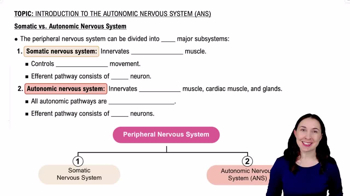
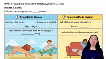
    Non-striated, spindle-shaped cells with single nucleus; contraction slower and sustained; controlled by autonomic nervous system.
  • Smooth muscle contraction regulation

    Ca2+ binds calmodulin, activating myosin light chain kinase (MLCK), which phosphorylates myosin light chains to enable contraction.
  • Cardiac muscle features

    Striated, branched fibers with single nucleus; connected by intercalated disks with gap junctions; involuntary control with pacemaker potentials.