Back

Muscle Tissue - Anatomy & Physiology

Control buttons has been changed to "navigation" mode.
1/27
  • Four properties of muscle tissue

    Contractility: ability to shorten or generate force.
    Excitability: ability to respond to a stimulus.
    Extensibility: ability to be stretched.
    Elasticity: ability to recoil after stretching.
  • Functions of muscle tissue

    Produces movement, opens and closes passageways, maintains posture and stabilizes joints, and generates heat during contraction.
  • Three muscle tissue types and control

    Skeletal: voluntary, striated, multinucleate.
    Cardiac: involuntary, striated, branched cells.
    Smooth: involuntary, nonstriated, fusiform cells.
  • Connective tissue layers of skeletal muscle

    Epimysium: surrounds entire muscle.
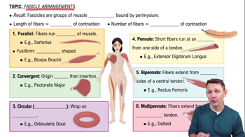
    Perimysium: surrounds fascicles.
    Endomysium: surrounds individual muscle fibers.
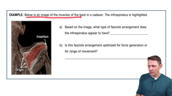
  • Difference between origin and insertion

    Origin is the less movable attachment; insertion is the more movable attachment of a muscle.
  • Organization levels from largest to smallest

    Muscle > Fascicle > Muscle fiber > Myofibril > Sarcomere > Myofilament.
  • Why skeletal muscle fibers are multinucleate

    Formed by fusion of many embryonic myoblasts, resulting in multiple nuclei per fiber.
  • Sarcomere definition and boundaries

    The contractile unit of a myofibril, extending from one Z disc to the next.
  • Thin and thick filament locations in sarcomere

    Thin filaments anchor at Z discs and extend inward; thick filaments are centered and overlap thin filaments.
  • Differences between A band, I band, and H zone

    A band: full length of thick filament.
    I band: only thin filaments.
    H zone: thick filaments only, no thin filament overlap.
  • Functions of titin in sarcomere

    Holds thick filaments in place, resists overstretching, and provides elastic recoil.
  • Components of a triad

    One T tubule flanked by two terminal cisterns of the sarcoplasmic reticulum.
  • Ion released by sarcoplasmic reticulum to initiate contraction

    Calcium ions.
  • Molecule that powers the sliding filament mechanism

    ATP.
  • Changes in sarcomere during contraction

    Z discs move closer, I bands shorten, H zone narrows or disappears, A band length remains constant.
  • Why 'sliding filament mechanism' is more accurate than filament shortening

    Filaments slide past each other without changing length, increasing overlap to shorten sarcomere.
  • Difference between concentric and eccentric contraction

    Concentric: muscle shortens while producing force.
    Eccentric: muscle lengthens while producing force.
  • Definition of neuromuscular junction

    The functional meeting point between a motor neuron ending and a skeletal muscle fiber.
  • Sequence from nerve impulse to muscle contraction

    Impulse reaches terminal bouton → acetylcholine released → sarcolemma excited → signal travels down T tubules → sarcoplasmic reticulum releases calcium → contraction initiated.
  • Definition of a motor unit

    One motor neuron and all the muscle fibers it innervates.
  • Characteristics of slow oxidative fibers

    High myoglobin, many mitochondria, slow contraction speed, and high fatigue resistance.
  • Characteristics of fast glycolytic fibers

    Low myoglobin, few mitochondria, large diameter, fast contraction, and rapid fatigue.
  • Two distinguishing features of cardiac muscle vs skeletal muscle

    Cardiac muscle cells branch and connect via intercalated discs; contract involuntarily and have 1-2 nuclei.
  • Differences between smooth and skeletal muscle

    Smooth muscle lacks striations, is involuntary, and found in hollow organs; skeletal muscle is striated, voluntary, and attached to bones.
  • Calcium sources for skeletal, cardiac, and smooth muscle

    Skeletal: sarcoplasmic reticulum only.
    Cardiac & smooth: sarcoplasmic reticulum and extracellular fluid.
  • Development sequence of skeletal muscle fibers

    Mesoderm cells divide → become myoblasts → fuse into multinucleate myotube → mature into skeletal muscle fiber.
  • Role of satellite cells in muscle growth

    Satellite cells are myoblast-like cells near fibers that fuse with existing fibers to aid growth and repair.