 Back
Muscle Tissue and Contraction - Anatomy & Physiology
 6:31

Terms in this set (23)
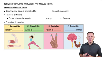
Excitability, contractility, extensibility, and elasticity.
Movement, posture maintenance, joint stabilization, and heat generation.
Epimysium surrounds the whole muscle, perimysium surrounds fascicles, and endomysium surrounds individual muscle fibers.
Actin filaments slide past myosin filaments, shortening the sarcomere and causing contraction.
Synapse where a motor neuron communicates with a skeletal muscle fiber using neurotransmitters.
Maintains resting membrane potential by pumping 3 Na+ out and 2 K+ in, keeping the cell polarized.
1. Excitation, 2. Excitation-contraction coupling, 3. Contraction (cross-bridge cycling), 4. Relaxation.
Myosin heads bind to actin, pivot to pull filaments, release with ATP, and reset to repeat contraction.
Provides energy for myosin head detachment and re-cocking during cross-bridge cycling.
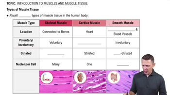
Muscle controlled without conscious effort, e.g., smooth and cardiac muscle.
Muscle controlled consciously, e.g., skeletal muscle.
Activation of cross-bridges to generate tension and shorten muscle fibers.
Acetylcholine (ACh) is released to stimulate muscle fiber contraction.
Small gap between the motor neuron and muscle fiber where neurotransmitters are released.
Ion channel that opens in response to changes in membrane potential.
Ion channel that opens when a specific chemical (neurotransmitter) binds to it.
State where the inside of the cell is negatively charged relative to the outside.
Loss of membrane potential as Na+ ions enter the cell, making it more positive.
Restoration of the resting membrane potential by K+ ions leaving the cell.
Thin filament protein that binds with myosin during contraction.
Thick filament protein with heads that form cross-bridges with actin.
Protein that blocks myosin-binding sites on actin when muscle is relaxed.
Protein that binds calcium and moves tropomyosin to expose binding sites on actin.