Skip to main content
Anatomy & Physiology
My Course
Learn
Exam Prep
AI Tutor
Study Guides
Textbook Solutions
Flashcards
Explore
Try the app
My Course
Learn
Exam Prep
AI Tutor
Study Guides
Textbook Solutions
Flashcards
Explore
Try the app
Back
Nervous System Anatomy & Physiology Study Guide
You can tap to flip the card.
Approximate number of neurons in the human brain
You can tap to flip the card.
👆
Approximate number of neurons in the human brain
About
20 billion
neurons.
Track progress
Control buttons has been changed to "navigation" mode.
1/100
Recommended videos
Guided course
1:10
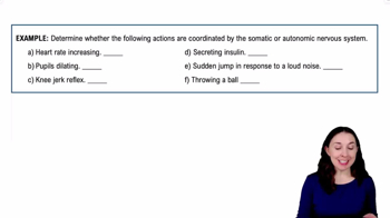
Introduction to the Autonomic Nervous System Example 1
8088
views
177
rank
Guided course
1:50
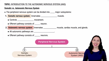
Somatic vs. Autonomic Nervous System
10722
views
215
rank
Guided course
3:10
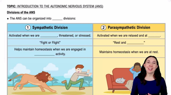
Divisions of the ANS
9149
views
142
rank
Terms in this set (100)
Hide definitions
Approximate number of neurons in the human brain
About
20 billion
neurons.
Structure separating the left and right cerebral hemispheres
The
longitudinal fissure
.
Grooves on the surface of the brain
Called
sulci
.
Ridges on the surface of the brain
Called
gyri
.
Brain region responsible for conscious thought and memory
The
cerebrum
.
Brain region that processes sensory information before the cerebrum
The
thalamus
.
Structure regulating emotions, autonomic functions, and hormone production
The
hypothalamus
.
Brain structure coordinating complex motor patterns and balance
The
cerebellum
.
Part of the brainstem controlling heart rate and breathing
The
medulla oblongata
.
Function of the pons
Relays signals between brain regions.
Another name for the midbrain
The
mesencephalon
.
Four fluid-filled cavities in the brain
Called
ventricles
.
Fluid filling the brain ventricles
Cerebrospinal fluid (CSF)
.
Structure producing cerebrospinal fluid
The
choroid plexus
.
Frequency of cerebrospinal fluid replacement
About every
8 hours
.
Three layers of the cranial meninges
Dura mater
,
arachnoid mater
, and
pia mater
.
Outermost meningeal layer
The
dura mater
.
Meningeal layer directly covering the brain surface
The
pia mater
.
Function of the blood-brain barrier
Protects the brain from harmful substances.
Artery supplying arterial blood to the brain
The
internal carotid arteries
.
Structure connecting the left and right cerebral hemispheres
The
corpus callosum
.
Lobe processing visual information
The
occipital lobe
.
Lobe processing auditory information
The
temporal lobe
.
Lobe controlling voluntary skeletal muscle movement
The
frontal lobe
.
Lobe processing touch and pressure sensations
The
parietal lobe
.
Function of the hippocampus
Memory formation.
Structure processing emotions such as fear and aggression
The
amygdala
.
Brain structure containing the pineal gland
The
epithalamus
.
Hormone produced by the pineal gland
Melatonin
.
Brain structure regulating body temperature and thirst
The
hypothalamus
.
Structure connecting hypothalamus to pituitary gland
The
infundibulum
.
Definition of ataxia
Loss of coordination.
Brain part monitoring proprioceptive input to coordinate movement
The
cerebellum
.
Brain area responsible for speech production
Broca’s area
.
Brain region responsible for understanding language
Wernicke’s area
.
Number of pairs of cranial nerves
12 pairs.
Numbering system for cranial nerves
Roman numerals.
Cranial nerve responsible for smell
Olfactory nerve (I).
Cranial nerve responsible for vision
Optic nerve (II).
Cranial nerve controlling most eye movements
Oculomotor nerve (III).
Cranial nerve controlling the superior oblique eye muscle
Trochlear nerve (IV).
Cranial nerve responsible for facial expressions
Facial nerve (VII).
Cranial nerve responsible for hearing and balance
Vestibulocochlear nerve (VIII).
Cranial nerve controlling swallowing and taste from the tongue
Glossopharyngeal nerve (IX).
Cranial nerve controlling organs in thoracic and abdominal cavities
Vagus nerve (X).
Cranial nerve controlling sternocleidomastoid and trapezius muscles
Accessory nerve (XI).
Cranial nerve controlling tongue movement
Hypoglossal nerve (XII).
Origin of cranial nerves
The brain.
Cranial nerve carrying parasympathetic signals to most visceral organs
Vagus nerve.
Type of nerve the facial nerve is
Mixed (sensory and motor).
Does the autonomic nervous system control voluntary or involuntary functions?
Involuntary functions.
Two divisions of the autonomic nervous system
Sympathetic and parasympathetic.
Division known as the 'fight-or-flight' system
Sympathetic division.
Division known as the 'rest-and-digest' system
Parasympathetic division.
Neurotransmitter released by most sympathetic postganglionic neurons
Norepinephrine.
Neurotransmitter released by parasympathetic neurons
Acetylcholine.
Spinal segments where sympathetic division originates
T1–L2.
Cranial nerves where parasympathetic division originates
III, VII, IX, X.
Cranial nerve providing most parasympathetic output
Vagus nerve.
Groups of neuron cell bodies outside the CNS
Ganglia.
Two neurons in an autonomic pathway
Preganglionic and postganglionic neurons.
Fiber connecting CNS to the ganglion
Preganglionic fiber.
Fiber connecting ganglion to the target organ
Postganglionic fiber.
Structures located on both sides of vertebral column in sympathetic division
Sympathetic chain ganglia.
Hormones released by the adrenal medulla
Epinephrine and norepinephrine.
Receptors responding to norepinephrine in sympathetic pathways
Adrenergic receptors.
Main function of the parasympathetic nervous system
Conserving energy and digestion.
Effect of parasympathetic stimulation on heart rate
Decreases heart rate.
Effect of parasympathetic stimulation on digestion
Increases digestion.
Effect of sympathetic stimulation on metabolism
Increases metabolism.
Type of reflex involving CNS in processing sensory input
Long reflex.
Reflex bypassing CNS and occurring in ganglia
Short reflex.
Parasympathetic stimulation is usually localized or widespread?
Localized.
Enzyme that breaks down acetylcholine
Acetylcholinesterase.
Sacral segments where parasympathetic fibers leave the spinal cord
S2–S4.
Difference between sensation and perception
Sensation is detection; perception is awareness.
Receptors that detect pain
Nociceptors.
Receptors detecting temperature changes
Thermoreceptors.
Receptors responding to mechanical distortion of membranes
Mechanoreceptors.
Receptors detecting chemical changes in body fluids
Chemoreceptors.
Receptors detecting body position
Proprioceptors.
Area monitored by a single receptor
Receptive field.
Term describing decreased sensitivity to constant stimuli
Adaptation.
The five special senses
Vision, hearing, taste, smell, equilibrium.
Location of olfactory receptors
Olfactory epithelium.
Structure carrying smell signals to the brain
Olfactory nerve.
Location of taste receptors
Tongue.
Structures containing taste receptors
Taste buds.
Typical lifespan of gustatory cells
7–10 days.
Inner ear structures detecting rotation of the head
Semicircular canals.
Structures detecting gravity and linear acceleration
Utricle and saccule.
Structure in cochlea containing hearing receptors
Organ of Corti.
Cranial nerve carrying hearing signals to the brain
Vestibulocochlear nerve (VIII).
Membrane vibrating when sound enters the ear
Tympanic membrane.
Three bones amplifying sound vibrations in the middle ear
Malleus, incus, stapes.
Part of the retina providing the sharpest vision
Fovea centralis.
Photoreceptors allowing vision in dim light
Rods.
Photoreceptors allowing color vision
Cones.
Structure of the eye focusing light onto the retina
Lens.
Condition when the retina separates from underlying tissue
Detached retina.
 BackBack
 1:10