 Back
Nervous System Fundamentals - Anatomy & Physiology
 04:36

Terms in this set (31)
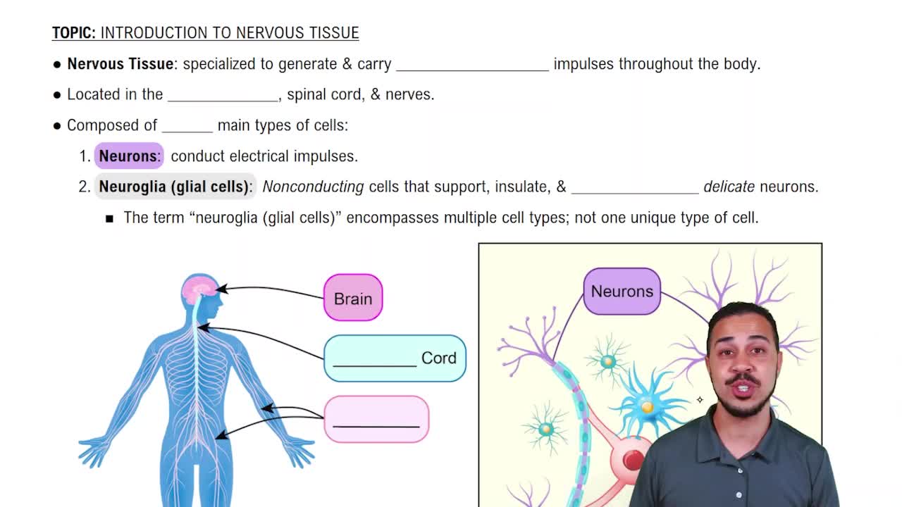
The nervous system is the major integrator of body function, working with the endocrine system to regulate homeostasis through rapid electrical signals and chemical messengers.
Neurons are excitable nerve cells that transmit electrical signals, have extreme longevity, are amitotic, and have a high metabolic rate requiring continuous O2 and glucose.
Neurons are classified by their processes: multipolar (most common), bipolar (rare), and unipolar (sensory neurons in dorsal root ganglia).
1. Cell body: biosynthetic center with nucleus.
2. Dendrites: receive graded potentials.
3. Axon: transmits impulses to other cells.
Dendrites are thin, highly branched extensions that serve as the receptive area for receiving graded potentials, providing a large surface area for information collection.
The axon hillock is the cone-shaped region of the soma where the threshold is reached to initiate an action potential.
The myelin sheath protects and electrically insulates axons, increasing impulse speed up to 150 times. In the PNS, it is formed by Schwann cells with gaps called Nodes of Ranvier.
1. Schwann cells: form myelin sheaths.
2. Satellite cells: cover neuron cell bodies in ganglia.
1. Astrocytes: maintain chemical environment.
2. Microglia: phagocytic cells.
3. Ependymal cells: line brain ventricles.
4. Oligodendrocytes: form CNS myelin sheaths.
A nerve is a cord-like organ containing axons, blood vessels, and connective tissue layers: endoneurium, perineurium, and epineurium.
1. Endoneurium: surrounds each axon.
2. Perineurium: surrounds fascicles.
3. Epineurium: surrounds entire nerve.
CNS: brain, spinal cord, interneurons, glial cells.
PNS: all nerves and ganglia outside CNS, including afferent and efferent fibers.
Voluntary system with afferent and efferent fibers from brain and spinal cord; uses acetylcholine as the only neurotransmitter.
Involuntary control of smooth, cardiac muscle, glands; includes sympathetic, parasympathetic, and enteric divisions; uses acetylcholine and norepinephrine.
Mobilizes body for 'fight or flight'; thoracolumbar origin; increases heart rate, blood pressure, blood sugar; neurotransmitters are acetylcholine and norepinephrine.
Rest and digest functions; craniosacral origin; decreases heart rate and blood pressure; increases digestion; uses acetylcholine as neurotransmitter.
Difference in electrical charge across the neuron membrane at rest, approximately -70mV, due to ionic concentration differences and membrane permeability.
1. Electrical attraction by fixed anions.
2. Greater permeability to K+ than Na+.
3. Active Na+/K+ ATPase pump maintaining ion gradients.
Depolarization: membrane potential becomes less negative (toward zero).
Hyperpolarization: membrane potential becomes more negative than resting.
Local changes in membrane potential that vary with stimulus intensity and decline over distance.
Summation of depolarizing graded potentials opens voltage-gated Na+ channels at the axon hillock once threshold (~-55mV) is reached.
Absolute refractory period: no new AP can be generated.
Relative refractory period: strong stimulus can trigger AP.
1. Axon diameter: larger diameter increases speed.
2. Myelin sheath: insulates and increases conduction rate.
Autoimmune attack on CNS myelin (oligodendrocytes) causing decreased conduction, leading to neuromuscular symptoms.
Group A: large, myelinated, 15-130 m/s.
Group B: intermediate, lightly myelinated, 3-15 m/s.
Group C: small, unmyelinated, ≤1 m/s.
Afferent: sensory, toward CNS.
Efferent: motor, away from CNS.
Interneurons: connect sensory and motor neurons within CNS.
Electrical synapses: direct ion flow, synchronize neurons.
Chemical synapses: neurotransmitter release across synaptic cleft.
1. Ca2+ gates open.
2. V-SNARE and T-SNARE bind.
3. Vesicle docks.
4. Neurotransmitter released.
5. Binds postsynaptic receptors.
6. Opens ion channels.
Excitatory: open Na+ and K+ channels, cause EPSPs.
Inhibitory: open K+ or Cl- channels, cause IPSPs.
1. Enzymatic degradation.
2. Reuptake into presynaptic terminal.
3. Diffusion away and systemic destruction.
Temporal summation: rapid impulses from one neuron.
Spatial summation: simultaneous impulses from multiple neurons.