 Back
BackNeural Tissue and Nervous System Anatomy & Physiology
 0:50
0:50
Terms in this set (28)
The CNS consists of the brain and spinal cord, responsible for processing and integrating information.
The PNS includes peripheral nerves and ganglia, connecting the CNS to limbs and organs.
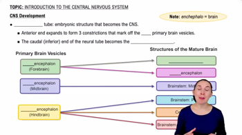
Process where the neural plate folds and fuses to form the neural tube, which develops into the CNS.
Cells at the border of the neural plate that migrate to form the PNS.
Neurons have a cell body (soma), dendrites (receive signals), and axon (sends signals).
Neurons have a resting voltage of about \(-70\,mV\), maintained by NaK ATPase.
When membrane potential reaches about \(-55\,mV\), voltage-gated Na+ channels open, triggering depolarization.
Includes depolarization, repolarization (K+ channels open), hyperpolarization, and refractory period.
Junction where neurotransmitters are released from axon terminals to bind receptors on target cells.
Sensory (afferent), motor (efferent), and interneurons (connect neurons within CNS).
Supportive cells in nervous tissue including astrocytes, oligodendrocytes, microglia, ependymal cells, Schwann cells, and satellite cells.
Most abundant CNS glial cells; maintain blood-brain barrier and support neuronal connections.
CNS glial cells that myelinate axons, increasing action potential speed.
PNS glial cells that myelinate axons, similar to oligodendrocytes but in the PNS.
CNS immune cells derived from blood monocytes; remove debris by phagocytosis.
Line brain ventricles and spinal cord central canal; produce and circulate cerebrospinal fluid (CSF).
Formed by astrocyte end-feet around blood vessels, regulating substance passage into the brain.
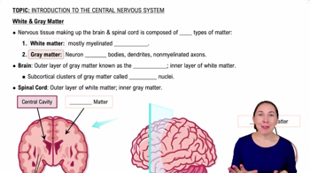
Grey matter contains neuron cell bodies and dendrites; white matter contains myelinated axons.
Grey matter forms a butterfly shape centrally; dorsal horn receives sensory input, ventral horn sends motor output.
Axons bundled with Schwann cells, surrounded by endoneurium, grouped into fascicles with perineurium, and covered by epineurium.
Gaps between myelinated segments on axons that facilitate rapid action potential propagation.
Clusters of neuronal cell bodies in the PNS, surrounded by connective tissue layers.
Characterized by tau protein tangles, amyloid beta plaques, neuronal death, and dysfunctional microglia.
Three connective tissue layers surrounding CNS: dura mater, arachnoid, and pia mater.
Outer folded grey matter layer of the brain with six layers of neurons, mainly pyramidal cells.
Action potential triggers Ca2+ influx, causing synaptic vesicles to release neurotransmitters into the synaptic cleft.
Multipolar, bipolar, unipolar, and anaxonic neurons differ in dendrite and axon number and arrangement.
Neuroepithelial progenitor cells differentiate into neuroblasts, neurons, and glial cells.