Skip to main content
Anatomy & Physiology
My Course
Learn
Exam Prep
AI Tutor
Study Guides
Textbook Solutions
Flashcards
Explore
Try the app
My Course
Learn
Exam Prep
AI Tutor
Study Guides
Textbook Solutions
Flashcards
Explore
Try the app
Back
Respiratory System - Anatomy & Physiology
You can tap to flip the card.
Organs of the respiratory system
You can tap to flip the card.
👆
Organs of the respiratory system
Nose, pharynx, larynx, trachea, bronchi, lungs (alveoli)
Track progress
Control buttons has been changed to "navigation" mode.
1/29
Recommended videos
4:28
Ventilation
7848
views
187
rank
1
comments
5:55
Ventilation and Respiration
11640
views
348
rank
1
comments
1:37
Introduction to Lung Physiology Example 1
7888
views
213
rank
1
comments
Terms in this set (29)
Hide definitions
Organs of the respiratory system
Nose, pharynx, larynx, trachea, bronchi, lungs (alveoli)
Where does gas exchange occur in the respiratory system?
Gas exchange occurs only in the
alveoli of the lungs
.
Upper vs Lower respiratory tract
Upper tract: passageways from nose to larynx. Lower tract: passageways from trachea to alveoli.
Functions of passageways to the lungs
They
purify, humidify, and warm
the incoming air.
Nasal cavity features
Contains
olfactory epithelium
, divided by nasal septum, lined with respiratory mucosa that moistens air and traps particles.
Role of conchae in the nasal cavity
Increase
surface area
and
air turbulence
to trap inhaled particles.
Paranasal sinuses functions
Lighten the skull, act as resonance chambers for speech, and produce mucus.
Pharynx regions
Nasopharynx
(behind nasal cavity),
oropharynx
(behind mouth),
laryngopharynx
(attached to larynx).
Function of the epiglottis
Protects the larynx opening by routing food to the esophagus and air to the trachea during swallowing.
Larynx main functions
Routes air and food into proper channels and plays a role in speech.
Structure of the trachea
4-inch tube with C-shaped hyaline cartilage rings, lined with ciliated mucosa and goblet cells producing mucus.
Differences between right and left main bronchi
Right bronchus is wider, shorter, and straighter than the left.
Lung lobes
Left lung has two lobes; right lung has three lobes.
Pleura and pleural fluid function
Pleura covers lungs and thoracic cavity; pleural fluid reduces friction and allows lung movement.
Respiratory zone structures
Includes respiratory bronchioles, alveolar ducts, alveolar sacs, and alveoli where gas exchange occurs.
Respiratory membrane composition
Formed by alveolar and capillary walls; allows gas diffusion between air and blood.
Role of alveolar macrophages
Remove bacteria, carbon particles, and debris to protect lungs.
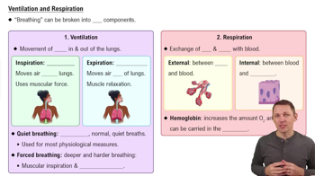
Four events of respiration
Pulmonary ventilation, external respiration, respiratory gas transport, internal respiration.
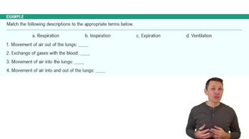
Pulmonary ventilation phases
Inspiration
(air into lungs) and
expiration
(air out of lungs).
Mechanics of inspiration
Diaphragm and external intercostals contract, increasing thoracic volume and decreasing pressure to draw air in.
Mechanics of expiration
Passive process where lung elasticity decreases volume and increases pressure to push air out.
Intrapleural pressure importance
Always negative to prevent lung collapse; equalizing with atmospheric pressure causes lung recoil.
Tidal volume (TV)
Amount of air moved in or out during normal quiet breathing, about 500 ml.
Inspiratory reserve volume (IRV)
Additional air forcibly inhaled beyond tidal volume, about 3,100 ml.
Expiratory reserve volume (ERV)
Additional air forcibly exhaled beyond tidal volume, about 1,200 ml.
Residual volume
Air remaining in lungs after forced expiration, about 1,200 ml, keeps alveoli inflated.
Vital capacity formula
Vital capacity = TV + IRV + ERV; total exchangeable air volume.
Dead space volume
Air in conducting zone that does not reach alveoli, about 150 ml.
Functional volume
Air that reaches the respiratory zone and participates in gas exchange, about 350 ml.
 BackBack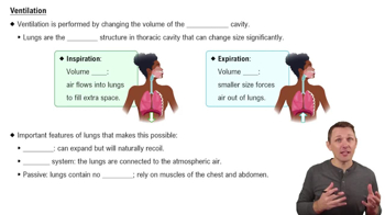
 4:28
4:28