Skip to main content
Anatomy & Physiology
My Course
Learn
Exam Prep
AI Tutor
Study Guides
Textbook Solutions
Flashcards
Explore
Try the app
My Course
Learn
Exam Prep
AI Tutor
Study Guides
Textbook Solutions
Flashcards
Explore
Try the app
Back
Respiratory System Anatomy & Physiology
You can tap to flip the card.
External portion of the nose
You can tap to flip the card.
👆
External portion of the nose
Supported by bone and hyaline cartilage, covered with muscle and skin, and lined with mucus membrane.
Track progress
Control buttons has been changed to "navigation" mode.
1/22
Recommended videos
4:28
Ventilation
7374
views
179
rank
1
comments
5:55
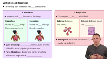
Ventilation and Respiration
11067
views
331
rank
1
comments
1:37
Introduction to Lung Physiology Example 1
7458
views
204
rank
1
comments
Terms in this set (22)
Hide definitions
External portion of the nose
Supported by bone and hyaline cartilage, covered with muscle and skin, and lined with mucus membrane.
Function of nasal vestibule
Contains hairs that remove dust particles from inhaled air.
Nasal septum composition
Wall of bone formed by the perpendicular plate of the ethmoid bone and the vomer, covered in mucus membrane.
Meatuses in the nasal cavity
Three groove-like passages formed by the superior, middle, and inferior nasal conchae projecting into the cavity.
Olfactory epithelium location
Mucus membrane lining the superior nasal conchae and adjacent septum containing olfactory receptors.
Pharynx divisions
Nasopharynx (respiratory only), oropharynx (conducts food and air), and laryngopharynx (conducts food and air).
Nasopharynx features
Superior portion, lined with cilia and mucus-producing cells, contains openings to internal nares and auditory tubes.
Larynx cartilage types
Includes thyroid, epiglottis, cricoid, arytenoid, corniculate, and cuneiform cartilages.
Function of the epiglottis
Covers the glottis during swallowing to prevent food from entering the larynx.
True vocal folds
Mucus membrane folds at the glottis that control the pitch of sound produced.
Structure of the tracheal wall
From deep to superficial: mucosa, submucosa, hyaline cartilage, and areolar connective tissue.
Tracheal cartilage rings
16-20 C-shaped rings of hyaline cartilage with open ends facing the esophagus to allow tracheal collapse during swallowing.
Carina of the trachea
Internal ridge of cartilage at the trachea's division into bronchi; very sensitive and triggers the cough reflex.
Primary bronchi characteristics
Formed by tracheal division; incomplete cartilage rings; right bronchus divides into 3, left into 2.
Bronchial tree branching order
Primary bronchi → secondary bronchi → tertiary bronchi → bronchioles → terminal bronchioles.
Changes in epithelium and cartilage in bronchioles
Cartilage rings replaced by plates; epithelium changes from ciliated simple columnar to nonciliated simple cuboidal.
Pleural membranes of the lungs
Parietal pleura (attached to thoracic wall), visceral pleura (covers lungs), and pleural cavity (contains lubricating fluid).
Lung surfaces
Base (inferior), apex (superior), costal surface (next to ribs), and medial surface (toward midline).
Bronchopulmonary segment
Lung tissue segment served by one tertiary bronchus; there are 10 in each lung.
Alveolar wall cell types
Type I alveolar cells (gas exchange), Type II alveolar cells (secrete surfactant), and alveolar macrophages (remove debris).
Function of surfactant secreted by Type II alveolar cells
Lowers surface tension of alveolar fluid to prevent alveoli collapse.
Alveolar/capillary membrane layers
Alveolar wall, epithelial basement membrane, capillary basement membrane, and endothelial cells of capillary.
 BackBack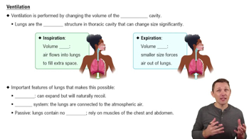
 4:28
4:28