 Back
Respiratory System - Anatomy & Physiology
 4:28

Terms in this set (31)
Oxygen and carbon dioxide exchange, protection of respiratory surfaces from dehydration, temperature changes and pathogens, sound production for communication, and detection of odors by olfactory receptors.
Upper respiratory system: nose, nasal cavity, sinuses, pharynx.
Lower respiratory system: larynx, trachea, bronchus, lungs (bronchioles and alveoli).
Mucous membrane lining nasal cavity, pharynx, larynx, trachea, bronchi, and bronchioles that traps debris and pathogens, protecting delicate exchange surfaces.
Provides airway for breathing, moistens and warms air, filters inhaled air, serves as resonating chamber for speech, and houses olfactory receptors.
Bony protrusions covered in thick mucus that increase surface area to warm, humidify, and filter incoming air.
Nasopharynx, oropharynx, and laryngopharynx; shared passageway for respiratory and digestive tracts.
Cartilaginous tube called the voice box containing vocal cords responsible for sound production and the glottis which controls air passage and phonation.
The vocal cords plus the opening between them in the larynx; controls airflow and sound production by opening and closing.
Air passes through the open glottis, vibrating the elastic vocal folds; pitch depends on diameter, length, and tension of vocal cords.
Thyroid cartilage (Adam's apple), cricoid cartilage, and epiglottis which prevents food from entering the respiratory tract during swallowing.
Tough, flexible tube supported by cartilaginous rings that conducts air to the lungs and prevents collapse or overexpansion during pressure changes.
A ridge of cartilage at the point where the trachea divides into left and right main bronchi.
Right lung has 3 lobes (superior, middle, inferior); left lung has 2 lobes (superior, inferior). Both lungs are protected by the pleural cavity with parietal and visceral pleura.
Trachea → primary (main) bronchi → secondary (lobar) bronchi → tertiary (segmental) bronchi → bronchioles → terminal bronchioles → respiratory bronchioles → alveoli.
Small air sacs covered by capillaries where gas exchange occurs between air and blood.
Alveolar cell layer, fused basement membrane, and capillary endothelial layer; allows efficient gas exchange.
Inflammation causes fluid leakage into alveoli, compromising the blood-air barrier and reducing gas exchange efficiency.
Sympathetic activation causes bronchodilation (enlargement), parasympathetic activation causes bronchoconstriction (narrowing).
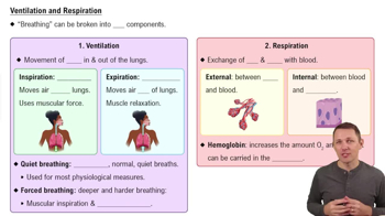
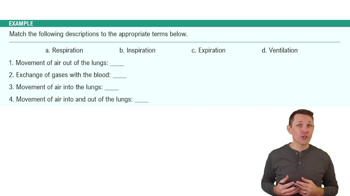
1. Breathing (pulmonary ventilation)
2. Gas diffusion across blood-air barrier
3. Transport of O2 and CO2 between alveoli and blood.
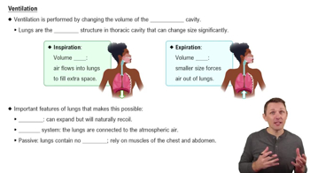
Diaphragm contracts downward and external intercostal muscles elevate the ribcage, increasing thoracic cavity volume to draw air in.
Diaphragm and external intercostal muscles relax, decreasing thoracic cavity volume and pushing air out; usually a passive process.
Adults: 12-18 breaths per minute; children: 18-20 breaths per minute.
Respiratory centers in the medulla oblongata stimulate diaphragm and external intercostal muscles to regulate breathing.
Monitor blood O2, CO2, and pH levels; increased CO2 triggers increased respiratory rate to expel excess CO2.
High blood pressure decreases respiratory rate; low blood pressure increases respiratory rate.
Prevent overexpansion of lungs during forced inhalation by triggering reflex inhibition of inspiration.
Coughing and sneezing involve apnea (temporary suspension of breathing) to expel irritants from airways.
Decreased lung capacity due to elastic tissue deterioration, arthritic rib joints restricting chest movement, and some emphysema is normal after age 50.
Chronic destruction of alveolar surfaces reduces gas exchange area, causing shortness of breath and reduced exercise tolerance; linked to smoking and aging.
Blockage of pulmonary artery by a traveling blood clot causing sudden shortness of breath and chest pain; often treated with blood thinners.
Hypoxia is low tissue oxygen levels; anoxia is complete oxygen deprivation causing rapid cell death.