 Back
Special Senses - Anatomy & Physiology
 5:05

Terms in this set (32)
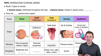
Smell, taste, sight, hearing, and equilibrium are the five special senses.
Large, complex sensory organs like the eye and ear, and localized clusters of receptors such as taste buds and olfactory epithelium.
About 70 percent of all sensory receptors are in the eyes.
Extrinsic eye muscles, eyelids, conjunctiva, and lacrimal apparatus.
They produce an oily secretion that lubricates the eye.
A membrane lining the eyelids and eyeball that secretes mucus to lubricate and keep the eye moist.
Includes the lacrimal gland and ducts; produces tears that cleanse, protect, moisten, and lubricate the eye.
Tears contain dilute salt solution, mucus, antibodies, and lysozyme (an enzyme that destroys bacteria).
Fibrous layer (outer), vascular layer (middle), and sensory layer (inner).
Sclera (white connective tissue) and cornea (transparent anterior portion).
It is a blood-rich nutritive layer containing pigment that prevents light scattering inside the eye.
The ciliary body (attached to the lens) and the iris (regulates light entering the eye).
Outer pigmented layer that absorbs light and inner neural layer containing photoreceptors (rods and cones).
Allow vision in dim light and peripheral vision, perceiving only gray tones.
Enable detailed color vision and are concentrated in the fovea centralis for sharpest vision.
It is where the optic nerve leaves the eyeball and contains no photoreceptors, so no image is detected there.
The anterior (aqueous) segment containing aqueous humor and the posterior (vitreous) segment containing vitreous humor.
It maintains intraocular pressure, provides nutrients to the lens and cornea, and is reabsorbed through the scleral venous sinus.
Through accommodation, the lens changes shape to focus light on the retina for objects less than 20 feet away.
Nearsightedness where distant objects appear blurry because the eyeball is too long and light focuses in front of the retina.
Farsightedness where near objects are blurry because the eyeball is too short or the lens is lazy, focusing light behind the retina.
Transmit and amplify vibrations from the tympanic membrane to the inner ear fluids via the malleus, incus, and stapes.
Cochlea, vestibule, and semicircular canals, filled with perilymph fluid.
The receptor organ for hearing located in the cochlear duct, containing hair cells that detect sound vibrations.
High-pitched sounds stimulate hair cells near the oval window; low-pitched sounds stimulate hair cells further along the cochlea.
Static equilibrium (head position) and dynamic equilibrium (angular or rotational movements).
Receptors in the vestibule that detect head position and help maintain head erectness by sending signals to the cerebellum.
Receptor in the semicircular canals that detects rotational head movements by hair cells covered with a gelatinous cupula.
Hindrance of sound vibration transmission through the external or middle ear.
Damage to nervous system structures involved in hearing, such as the cochlear nerve or auditory cortex.
In the roof of the nasal cavity; chemicals must dissolve in mucus to stimulate olfactory receptor neurons with cilia called olfactory hairs.
Sweet, sour, bitter, salty, and umami (savory taste from glutamate).