What is the extent of the spinal cord in the vertebral column?
The spinal cord extends from the foramen magnum to the inferior border of L1 vertebra.
Name the enlargements of the spinal cord and their significance.
Cervical enlargement and lumbosacral enlargement; they are regions where limbs are serviced.
How many spinal cord segments are there and what structures are associated with each segment?
There are 31 spinal cord segments, each associated with dorsal root ganglia, dorsal root, ventral root, and a spinal nerve.
What are the three layers of spinal meninges and their functions?
Dura mater (tough outer layer), arachnoid mater (middle layer with CSF-filled subarachnoid space), pia mater (innermost layer with blood vessels).
What is the epidural space and where is it located?
The epidural space is between the dura mater and the vertebral bones, containing adipose tissue and blood vessels.
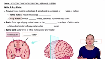
Describe the organization of gray matter in the spinal cord.
Gray matter is H-shaped, contains neuron cell bodies, organized into posterior (sensory), anterior (somatic motor), and lateral (visceral motor) horns.
What is the functional difference between the posterior and anterior gray horns?
Posterior gray horns contain somatic and visceral sensory nuclei; anterior gray horns contain somatic motor nuclei.
How is white matter in the spinal cord organized?
White matter is organized into columns (funiculi) containing tracts that are either ascending (sensory) or descending (motor).
How many pairs of spinal nerves are there and how are they named?
31 pairs: 8 cervical, 12 thoracic, 5 lumbar, 5 sacral, 1 coccygeal; cervical nerves named for vertebra below, thoracic and below named for vertebra above.
What are the three connective tissue layers of a spinal nerve?
What are the main branches of a spinal nerve after the dorsal and ventral roots fuse?
Dorsal ramus (serves skin and muscles of back), ventral ramus (serves ventrolateral body and limbs), and in T1-L2, white and gray rami communicantes.
What is a nerve plexus and which major plexuses exist?
A nerve plexus is a network of converging and branching ventral rami; major plexuses are cervical, brachial, lumbar, and sacral.
What nerves arise from the brachial plexus and what do they innervate?
Musculocutaneous, axillary, median, radial, and ulnar nerves; they innervate muscles of the pectoral girdle and upper limbs.
What nerves arise from the lumbar and sacral plexuses?
Lumbar plexus: femoral, obturator, iliohypogastric, ilio-inguinal nerves; sacral plexus: sciatic, pudendal, superior and inferior gluteal nerves.
What is a dermatome?
A dermatome is an area of skin innervated by sensory fibers from a single spinal nerve.
Define a reflex and the components of a reflex arc.
A reflex is an immediate involuntary motor response to a stimulus; reflex arc includes receptor, sensory neuron, integration center, motor neuron, and effector.
What is the difference between monosynaptic and polysynaptic reflexes?
Monosynaptic reflexes have a direct sensory-motor neuron synapse (e.g., stretch reflex); polysynaptic reflexes involve interneurons and multiple synapses.
What clinical conditions are associated with peripheral nerve palsies?
Conditions include brachial plexus birth palsy (Erb's palsy), pressure palsies, carpal tunnel syndrome, and sciatica.
What is a lumbar puncture and where is the needle inserted?
A lumbar puncture samples cerebrospinal fluid; needle is inserted into the subarachnoid space between L2 and sacrum.
What is an epidural block and its effect?
Injection of anesthetic into the epidural space; blocks sensory nerves in the area, often sparing motor function.
 Back

 0:50