 Back
Urinary System and Fluid Balance - Anatomy & Physiology

 0:54

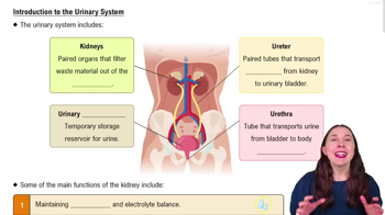
Terms in this set (42)
Filters blood to remove waste, regulates fluid and electrolyte balance, controls blood pressure, and maintains acid-base homeostasis.
Located retroperitoneally; covered by a renal capsule, surrounded by adipose tissue and renal fascia.
Ureters transport urine from kidneys to bladder; bladder stores urine; urethra conducts urine out of the body.
Male urethra is longer and serves both urinary and reproductive systems; female urethra is shorter and only for urine excretion.
Cortex (outer layer), medulla (inner layer), renal pyramids, and major blood vessels like the renal artery and renal vein.
Blood enters via afferent arteriole → glomerulus → efferent arteriole → peritubular capillaries or vasa recta → renal vein.
Includes the glomerulus (capillary network) and Bowman's capsule (surrounding capsule).
Cortical nephrons have short loops of Henle; juxtamedullary nephrons have long loops important for urine concentration.
Glomerular capillaries filter blood; peritubular capillaries surround tubules for reabsorption and secretion.
Includes macula densa, juxtaglomerular cells, and extraglomerular mesangial cells; regulates blood pressure and filtration.
From renal corpuscle → proximal tubule → loop of Henle → distal tubule → collecting duct → ureter → bladder → urethra.
Filtration in glomerulus, reabsorption mainly in tubules, and secretion in tubules.
Consists of fenestrated endothelium, basement membrane, and podocyte filtration slits.
Constriction of efferent arteriole and dilation of afferent arteriole maintain high pressure for filtration.
Hydrostatic pressure pushes fluid out; colloid osmotic pressure pulls fluid in; balance determines filtration rate.
Average GFR is about 125 mL/min in healthy adults.
Blood pressure, sodium levels, and hormones like renin-angiotensin affect GFR.
Include active transport, osmosis, and facilitated diffusion depending on nephron segment.
Protein channels that facilitate water reabsorption across nephron tubule membranes.
Includes Na+, K+, Ca2+, and glucose at various nephron segments.
Loop of Henle and vasa recta create a gradient to concentrate urine by reabsorbing water and solutes.
Occurs mainly in the proximal tubule, distal tubule, and collecting duct.
Reabsorption moves solutes from tubule to blood; secretion moves solutes from blood to tubule; energy use varies.
Filtration, reabsorption, and secretion rates determine solute excretion like sodium.
Intrinsic mechanisms that regulate afferent arteriole diameter to control GFR and urine volume.
Renin-angiotensin constricts arterioles; sympathetic activity reduces GFR during stress.
Renin-angiotensin, aldosterone, and ADH regulate sodium and water reabsorption affecting urine concentration.
Ureters transport urine; bladder stores urine; urethra expels urine.
Involves involuntary spinal reflex and voluntary control via the external urethral sphincter.
Include intracellular fluid and extracellular fluid (interstitial fluid and plasma).
Substances that dissociate into ions and conduct electricity in solution.
Sodium regulates fluid balance; potassium controls membrane potential; chloride balances charge; calcium supports bones and muscle function.
Electrolytes determine osmotic pressure by controlling water movement between compartments.
Acid releases H+; base accepts H+; pH measures H+ concentration; buffers resist pH changes.
Between 7.35 and 7.45.
Maintains pH by reversible reaction between H2CO3 and HCO3- to neutralize acids and bases.
PCO2: 35-45 mmHg; HCO3-: 22-26 mEq/L; both influence blood pH.
Controls CO2 levels; hypoventilation increases CO2 and lowers pH; hyperventilation decreases CO2 and raises pH.
Secretes H+ and reabsorbs or produces HCO3- to regulate blood pH.
Respiratory or renal adjustments restore pH by altering CO2 or HCO3- levels.
Cardiovascular, endocrine, and urinary systems coordinate to regulate volume and pH.
Disruptions like hypoventilation, diarrhea, or kidney dysfunction affect acid-base balance.