 Back
BackUrinary System and Kidney Function - Anatomy & Physiology

 0:54
0:54
Terms in this set (29)
Regulate osmolality, water volume, ion concentration, blood cell formation (erythropoietin), blood pressure (renin), acid-base balance, filter metabolic wastes, activate Vitamin D, and perform gluconeogenesis during fasting.
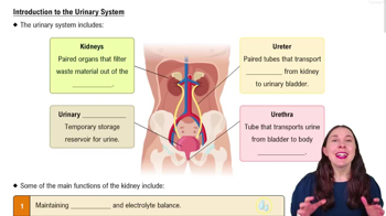
Kidneys, ureters, urinary bladder, and urethra.
Opening in the kidney for the ureter, blood vessels, lymphatics, and nerves.
Renal fascia (superficial, attachment), perirenal capsule (middle, trauma protection), fibrous capsule (deep, infection protection).
Renal cortex (outermost), renal medulla (middle, contains pyramids and columns), renal pelvis (innermost, continuous with ureter).
Nephron, consisting of renal corpuscle and renal tubules.
Cortical nephrons mostly in cortex with short loops; juxtamedullary nephrons near cortex-medulla junction with long loops and vasa recta.
First capillary bed in nephron; filters blood to form filtrate.
Afferent arterioles carry blood to glomerulus; efferent arterioles carry blood away to peritubular capillaries or vasa recta.
Glomerular filtration, tubular reabsorption, and tubular secretion.
Filtrate is blood plasma without proteins; urine is filtrate with unneeded substances like excess salts and wastes.
Endothelial fenestrations, basement membrane, and filtration slits between podocytes.
Hydrostatic pressure of glomerular capillaries (HPgc) pushes water and solutes out of blood.
Hydrostatic pressure in capsular space (HPcs) and osmotic pressure in glomerular capillaries (OPgc).
\(\text{NFP} = HP_{gc} - (HP_{cs} + OP_{gc})\)
120-125 ml/min; volume of filtrate formed per minute by all glomeruli.
Myogenic response (arteriole constriction/dilation) and tubular glomerular feedback via juxtaglomerular complex.
Monitor NaCl concentration in filtrate and signal afferent arteriole to constrict or dilate to regulate GFR.
Renin converts angiotensinogen to angiotensin I; ACE converts to angiotensin II which increases BP by vasoconstriction, aldosterone release, ADH release, and thirst stimulation.
Increases aquaporins in collecting ducts, promoting water reabsorption and reducing urine output.
Na+/K+ pump moves Na+ out of tubule cells against gradient, creating electrochemical gradient for secondary transport.
Symporters move Na+ and other molecules in same direction; antiporters move Na+ and H+ in opposite directions.
Water moves from low solute to high solute concentration areas, mainly through aquaporins by facilitated diffusion.
Creates concentration gradient in medulla by NaCl movement in ascending loop and water movement in descending loop to concentrate urine.
Countercurrent exchanger that preserves medullary gradient and removes reabsorbed water without washing out solutes.
Urea is reabsorbed and recycled in medulla to maintain high solute concentration and promote water reabsorption when ADH is present.
Removes substances not filtered at glomerulus, disposes of drugs, wastes, excess K+, and helps regulate blood pH.
Increases Na+/K+ pump activity, promoting sodium reabsorption and increasing blood pressure via RAA system.
Internal urethral sphincter is involuntary smooth muscle; external urethral sphincter is voluntary skeletal muscle.